Biomarker assay of neurological condition

a biomarker and neurological technology, applied in the field of neurological condition determination, can solve the problems of brain damage prospect, diagnostic limitations, and high cost of spectroscopic imaging, and achieve the effect of reducing the cost and time of spectroscopic imaging

Inactive Publication Date: 2017-11-02
BANYAN BIOMARKERS INC
View PDF3 Cites 3 Cited by
  • Summary
  • Abstract
  • Description
  • Claims
  • Application Information

AI Technical Summary

Problems solved by technology
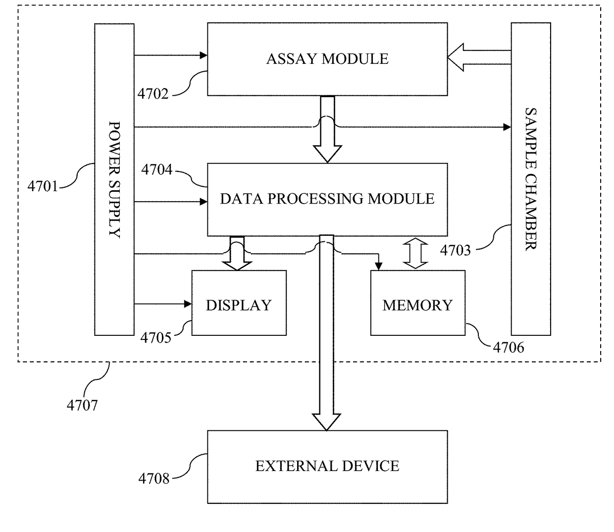
Traumatic, ischemic, and neurotoxic chemical insult, along with generic disorders, all present the prospect of brain damage.
While the diagnosis of severe forms of each of these causes of brain damage is straightforward through clinical response testing and computed tomography (CT) and magnetic resonance imaging (MRI) testing, these diagnostics have their limitations in that spectroscopic imaging is both costly and time consuming while clinical response testing of incapacitated individuals is of limited value and often precludes a nuanced diagnosis.
Additionally, owing to the limitations of existing diagnostics, situations under which a subject experiences a stress to their neurological condition such that the subject often is unaware that damage has occurred or seek treatment as the subtle symptoms often quickly resolve.
The lack of treatment of these mild to moderate challenges to neurologic condition of a subject can have a cumulative effect or subsequently result in a severe brain damage event which in either case has a poor clinical prognosis.

Method used


Image

  • Biomarker assay of neurological condition
  • Biomarker assay of neurological condition
  • Biomarker assay of neurological condition

Examples


example 1

Materials for Biomarker Analyses

[0179]Illustrative reagents used in performing the subject invention include Sodium bicarbonate (Sigma Cat #: C-3041), blocking buffer (Startingblock T20-TBS) (Pierce Cat#: 37543), Tris buffered saline with Tween 20 (TBST; Sigma Cat #: T-9039). Phosphate buffered saline (PBS; Sigma Cat #: P-3813); Tween 20 (Sigma Cat #: P5927); Ultra TMB ELISA (Pierce Cat #: 34028); and Nunc maxisorp ELISA plates (Fisher). Monoclonal and polyclonal GFAP and UCH-L1 antibodies are made in-house or are obtained from Santa Cruz Biotechnology, Santa Cruz, Calif. Antibodies directed to □-II spectrin and breakdown products as well as to MAP2 are available from Santa Cruz Biotechnology, Santa Cruz, Calif. Labels for antibodies of numerous subtypes are available from Invitrogen, Corp., Carlsbad, Calif. Protein concentrations in biological samples are determined using bicinchoninic acid microprotein assays (Pierce Inc., Rockford, Ill., USA) with albumin standards. All other nec...

example 2

Biomarker Assay Development

[0180]Anti-biomarker specific rabbit polyclonal antibody and monoclonal antibodies are produced in the laboratory. To determine reactivity specificity of the antibodies to detect a target biomarker a known quantity of isolated or partially isolated biomarker is analyzed or a tissue panel is probed by western blot. An indirect ELISA is used with the recombinant biomarker protein attached to the ELISA plate to determine optimal concentration of the antibodies used in the assay. Microplate wells are coated with rabbit polyclonal anti-human biomarker antibody. After determining the concentration of rabbit anti-human biomarker antibody for a maximum signal, the lower detection limit of the indirect ELISA for each antibody is determined. An appropriate diluted sample is incubated with a rabbit polyclonal antihuman biomarker antibody for 2 hours and then washed. Biotin labeled monoclonal anti-human biomarker antibody is then added and incubated with captured biom...

example 3

In Vivo Model of TBI Injury Model

[0181]A controlled cortical impact (CCI) device is used to model TBI on rats as previously described (Pike et al, 1998). Adult male (280-300 g) Sprague-Dawley rats (Harlan: Indianapolis, Ind.) are anesthetized with 4% isoflurane in a carrier gas of 1:1 O2 / N2O (4 min.) and maintained in 2.5% isoflurane in the same carrier gas. Core body temperature is monitored continuously by a rectal thermistor probe and maintained at 37±1° C. by placing an adjustable temperature controlled heating pad beneath the rats. Animals are mounted in a stereotactic frame in a prone position and secured by ear and incisor bars. Following a midline cranial incision and reflection of the soft tissues, a unilateral (ipsilateral to site of impact) craniotomy (7 mm diameter) is performed adjacent to the central suture, midway between bregma and lambda. The dura mater is kept intact over the cortex. Brain trauma is produced by impacting the right (ipsilateral) cortex with a 5 mm d...


PUM


Abstract

A robust, quantitative, and reproducible process and assay for diagnosis of a neurological condition in a subject is provided. With measurement of one or more autoantibodies to biomarkers in a biological fluid such as CSF or serum, the extent of neurological damage in a subject with an abnormal neurological condition is determined and subtypes thereof or tissue types subjected to damage are discerned.

Description

CROSS REFERENCE TO RELATED APPLICATIONS[0001]This application is a continuation in part application of U.S. patent application Ser. No. 13 / 379,164 filed Dec. 19, 2011 that in turn claims priority of Patent Cooperation Treaty Application Serial No. PCT / US2010 / 039335 filed Jun. 21, 2010 which claims priority to U.S. Provisional Application No. 61 / 218,727 filed Jun. 19, 2009 and U.S. Provisional Application No. 61 / 345,188 filed May 17, 2010; and Patent Cooperation Treaty Application Serial No. PCT / US2011 / 040998 filed Jun. 17, 2011, which in turn claims priority benefit of U.S. Provisional Application Ser. No. 61 / 355,779, filed Jun. 17, 2010 and U.S. Provisional Application Ser. No. 61 / 476,158, filed Apr. 15, 2011; the contents of each of which are incorporated herein by reference in their entirety.FIELD OF THE INVENTION[0002]The present invention relates in general to determination of a neurological condition of an individual and in particular to measuring a quantity of a neuropredicti...

Claims


Application Information

Patent Timeline
Patent Type & Authority Applications(United States)
IPC IPC(8): G01N33/68
CPCG01N33/6896G01N2800/28
Inventor WANG, KEVIN KA-WANGZHANG, ZHIQUNLIU, MING CHENGHAYES, RONALD L.
Owner BANYAN BIOMARKERS INC
Why Eureka
Try Eureka