Method for automatic visual annotation of radiological images from patient clinical data

a radiological image and clinical data technology, applied in the field of medical image annotation, can solve the problem of presenting a resource challenge in the annotation of these databases

Inactive Publication Date: 2018-03-01
IBM CORP
View PDF1 Cites 5 Cited by
  • Summary
  • Abstract
  • Description
  • Claims
  • Application Information

AI Technical Summary

Benefits of technology

[0015]The system, in an optional embodiment, may further comprise an object matching engine configured to receive an image and the output of the textual analysis engine and generate a set of semantic descriptions for visual candidates and a matching e

Problems solved by technology

Annotation of these databases provides a resource challenge, as the number

Method used


Image

  • Method for automatic visual annotation of radiological images from patient clinical data
  • Method for automatic visual annotation of radiological images from patient clinical data
  • Method for automatic visual annotation of radiological images from patient clinical data

Examples


Embodiment Construction

[0026]In the following detailed description of the preferred embodiments, reference is made to the accompanying drawings, which form a part hereof, and within which are shown by way of illustration specific embodiments by which the invention may be practiced. It is to be understood that other embodiments may be utilized and structural changes may be made without departing from the scope of the invention. The following detailed description is therefore not to be taken in a limiting sense, and the scope of the present disclosure is defined by the appended claims and their equivalents.

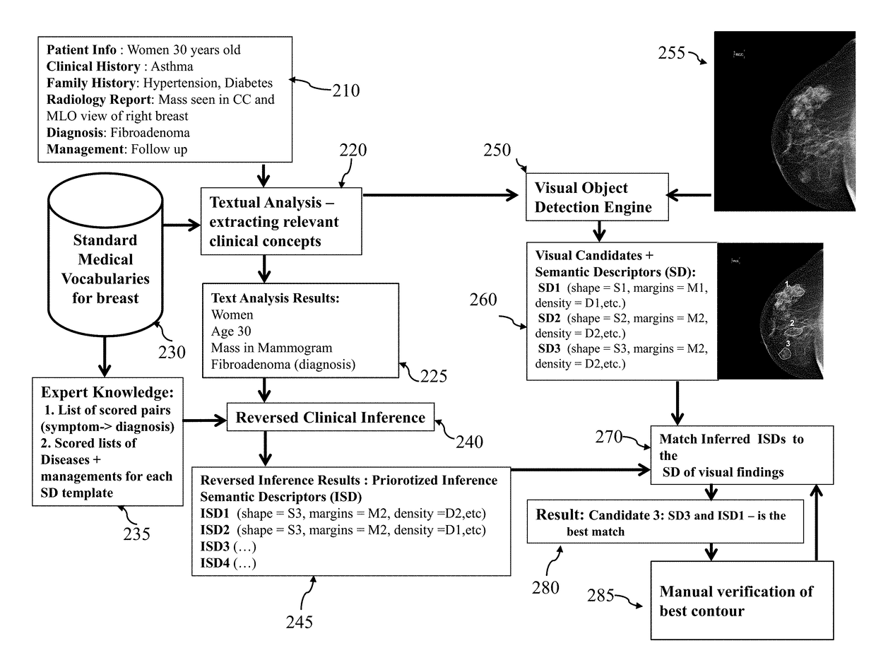
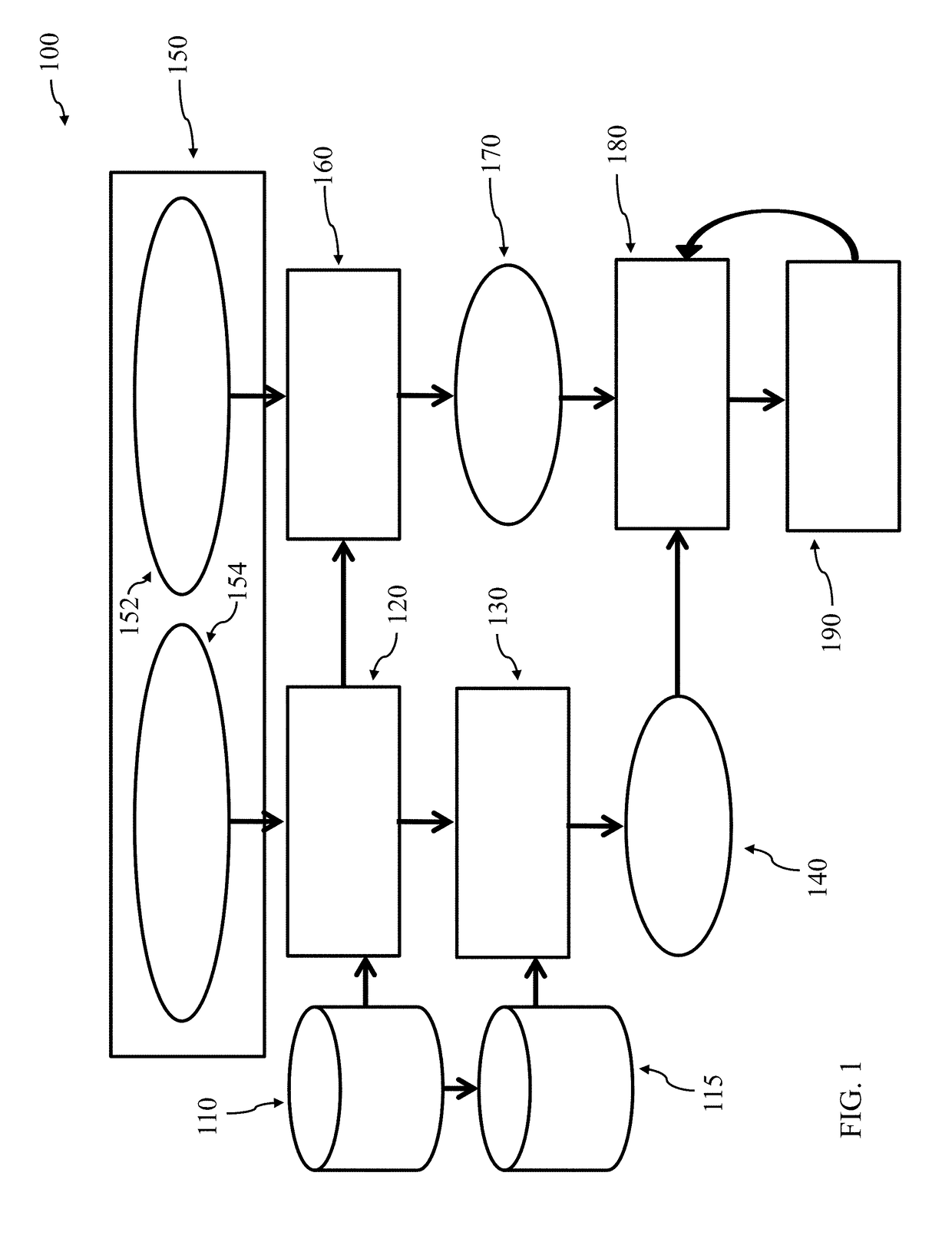
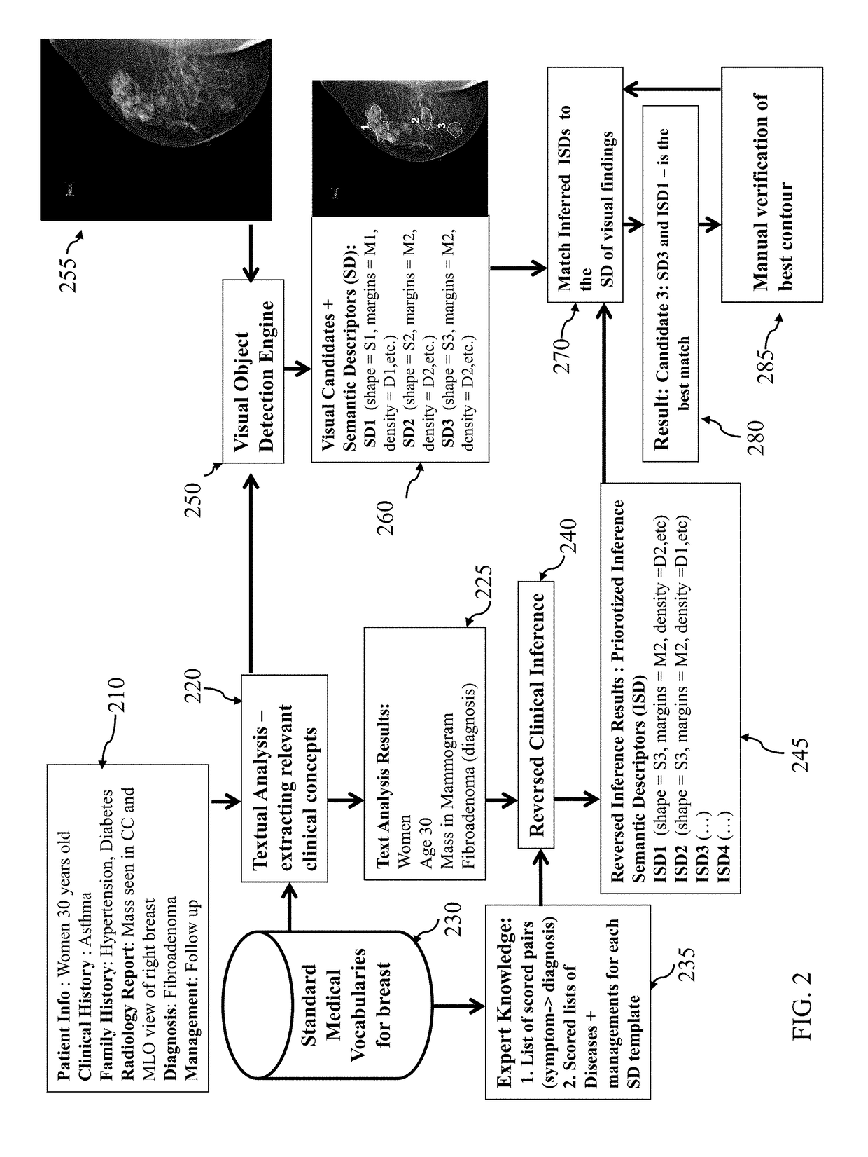
[0027]FIG. 1 illustrates a flow diagram 100 of the various components and data sources, according to an embodiment of the present invention. The system initially receives a patient case 150. The patient case can be received over a computer network interface or other data interface and can comprise an electronic data file or stream. This patient case 150 contains both a set of textual data 154 and a set of...


PUM


Abstract

Presented herein are methods, systems, devices, and computer-readable media for image annotation for medical procedures. The system operates in a parallel manner. In one flow, the system starts from clinical terms and image and applies image detection module in order to get visual candidates for related radiological finding and provide them with semantic descriptors. In the second (parallel) flow, the system produces a list of prioritized semantic descriptors (with probabilities). The second flow is done by application of a reverse inference algorithm that uses clinical terms and expert clinical knowledge. The results of both flows combined by matching module for detection the best candidate and with limited user input images can be annotated. The clinical terms are extracted from clinical documents by textual analysis.

Description

FIELD OF TECHNOLOGY[0001]The present invention relates to the technical field of medical image annotation. More particularly, the present invention is in the field of automated image annotation using reverse inference.BACKGROUND OF THE INVENTION[0002]Medical imaging has grown over the past decades to become an essential component of diagnoses and treatment. This field has seen significant developments in applications for computer-assisted diagnostics and image-guided medical procedures. These advances are tied, in part, to technical and scientific improvements in imaging. For example, some of the early work in this field in the late 1980s provided for medical image shape detection. These were some of the building blocks of systems developed in the mid-1990s and thereafter, such as image-guided surgery systems. These diagnostics systems aid medical practitioners in identifying diseases, and image-guided surgery makes use of imaging to aid a surgeon in performing more effective and ac...

Claims


Application Information

Patent Timeline
IPC IPC(8): G06F19/00G06F17/30G06F17/24G06N5/02G06N5/04G06N99/00G06N7/00G06N20/00
CPCG06F19/321G06F19/345G06F17/30401G06F17/241G16H50/20G06N5/04G06N99/005G06N7/005G06N5/022G16H30/40G16H40/60G16H50/70G16H70/60G06F40/169G06N20/00G06F16/243
Inventor BARKAN, ELLAKISILEV, PAVELWALACH, EUGENE
Owner IBM CORP
Why Eureka
Try Eureka