Automatic Liver Segmentation Using Adversarial Image-to-Image Network

an adversarial image-to-image network and liver segmentation technology, applied in image data processing, instruments, computing, etc., can solve the problems of inability to provide accurate liver segmentation and high difficulty in automatic liver segmentation in medical images

Active Publication Date: 2018-09-13
SIEMENS HEALTHCARE GMBH
View PDF10 Cites 108 Cited by
  • Summary
  • Abstract
  • Description
  • Claims
  • Application Information

AI Technical Summary

Problems solved by technology

However, automatic liver segmentation in medical images is a highly challenging task due to complex background, fuzzy boundaries, and varying appearance of the liver in medical images.
However, due to the challe

Method used


Image

  • Automatic Liver Segmentation Using Adversarial Image-to-Image Network
  • Automatic Liver Segmentation Using Adversarial Image-to-Image Network
  • Automatic Liver Segmentation Using Adversarial Image-to-Image Network

Examples


Embodiment Construction

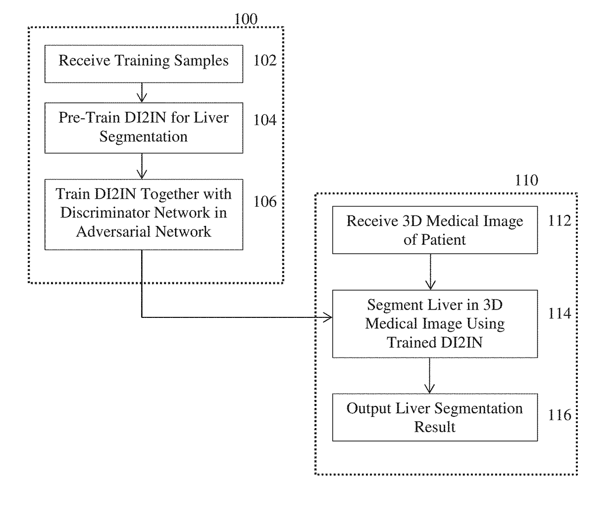
[0018]The present invention relates to a method and system for automated computer-based liver segmentation in 3D medical images. Embodiments of the present invention are described herein to give a visual understanding of automated liver segmentation method. A digital image is often composed of digital representations of one or more objects (or shapes). The digital representation of an object is often described herein in terms of identifying and manipulating the objects. Such manipulations are virtual manipulations accomplished in the memory or other circuitry / hardware of a computer system. Accordingly, is to be understood that embodiments of the present invention may be performed within a computer system using data stored within the computer system.

[0019]Various methods have been proposed for computer-based automatic liver segmentation from 3D CT scans. Such methods can be generally categorized into non-learning-based and learning-based approaches. Non-learning-based approaches usua...


PUM


Abstract

A method and apparatus for automated liver segmentation in a 3D medical image of a patient is disclosed. A 3D medical image, such as a 3D computed tomography (CT) volume, of a patient is received. The 3D medical image of the patient is input to a trained deep image-to-image network. The trained deep image-to-image network is trained in an adversarial network together with a discriminative network that distinguishes between predicted liver segmentation masks generated by the deep image-to-image network from input training volumes and ground truth liver segmentation masks. A liver segmentation mask defining a segmented liver region in the 3D medical image of the patient is generated using the trained deep image-to-image network.

Description

[0001]This application claims the benefit of U.S. Provisional Application No. 62 / 468,400, filed Mar. 8, 2017, the disclosure of which is herein incorporated by reference.BACKGROUND OF THE INVENTION[0002]The present invention relates to automatic liver segmentation in medical images, and more particularly, to deep-learning based automatic liver segmentation in 3D medical images.[0003]Accurate liver segmentation in three dimensional (3D) medical images, such as computed tomography (CT) or magnetic resonance (MR) images, is important in many clinical applications, such as pathological diagnosis of hepatic diseases, surgical planning, and postoperative assessment. However, automatic liver segmentation in medical images is a highly challenging task due to complex background, fuzzy boundaries, and varying appearance of the liver in medical images.[0004]Various methods have been proposed for computer-based automatic liver segmentation from 3D CT scans. Such methods can be generally categor...

Claims


Application Information

Patent Timeline
IPC IPC(8): G06T7/11G06T7/187
CPCG06T7/11G06T7/187G06T2207/20081G06T2207/30056G06T2207/10081G06T2207/20084G06T7/0012G06T2207/10088G06T2207/10104G06T2207/20076G06T7/143
Inventor YANG, DONGXU, DAGUANGZHOU, SHAOHUA KEVINGEORGESCU, BOGDANCHEN, MINGQINGCOMANICIU, DORIN
Owner SIEMENS HEALTHCARE GMBH
Why Eureka
Try Eureka