Quantum Mechanical/X-Ray Crystallography Diagnostic for Proteins

a technology of x-ray crystallography and quantum mechanical, applied in the direction of material analysis using wave/particle radiation, instruments, molecular structure, etc., can solve the problems of insufficient information about epitopes, protonation or solvent effects, x-ray crystallography, etc., to accurately assess protein conformation, protonation and solvent effects, useful and reliable

Active Publication Date: 2019-01-24
QUANTUMBIO INC
View PDF0 Cites 0 Cited by
  • Summary
  • Abstract
  • Description
  • Claims
  • Application Information

AI Technical Summary

Benefits of technology

[0007]In order to meet this need, the present invention is a diagnostic which bolsters x-ray crystallography with the addition of semi-empirical quantum mechanics analysis to give a verifiable physical and biochemical assessment of a protein or proteins. Even though neutron diffraction is too labor intensive to use on a regular basis for protein testing generally, for the purpose of verifying this invention neutron diffraction has confirmed for three examples that the present quantum mechanical x-ray crystallography diagnostic accurately assesses protein conformation, protonation and solvent effects. Therefore, neutron diffraction has already confirmed that the present tool is a useful and reliable diagnostic of protein structure and reactivity in vivo as well as in vitro. The present quantum mechanical x-ray crystallography diagnostic takes x-ray crystallographic data collected from a protein of interest and overlays (bolsters) the x-ray crystallography with a semi-empirical quantum mechanical set of identification steps as follows, to give a real world diagnostic of the status, conformation and reactivity of the protein in question. Specifically, the overlay in part takes the form of what we call a scoring event. In other words, the present invention includes a novel scoring method, called XModeScore, which (1) enumerates the possible protomeric / tautomeric modes, and thereafter (2) uses X-ray crystallographically to refine each mode specifically by using the semiempirical quantum mechanics (PM6) Hamiltonian, and subsequently (3) we score each mode using a combination of energetic strain (or ligand strain) and rigorous statistical analysis of the difference electron density distribution. By performing these three steps as what we call XModeScore, we are able consistently to distinguish the correct bound protomeric / tautomeric modes based on routine X-ray data—even at x-ray crystallography resolutions that those skilled in the art would consider to be “low.” As mentioned above, we have confirmed these bolstered x-ray crystallography results with results obtained from much more expensive and laborious neutron diffraction studies for three different examples: tautomerism in the acetazolamide ligand of human carbonic anhydrase II (PDB 3HS4 and 4K0S); tautomerism in the 8HX ligand of urate oxidase (PDB 4N9S and 4N9M); and protonation states of the catalytic aspartic acid found within the active site of an aspartic protease (PDB 2JJJ). In each case, XModeScore conducted with the X-ray crystallography diffraction data identified the correct protonation state as defined by the neutron diffraction data, thus corroborating the real-world efficacy of the present invention.

Problems solved by technology

Theoretical models alone are virtually impossible to use, given the unpredictable conformations of proteins or their protonation and solvent effects, that is, the chemical as well as the physical behavior of the protein or proteins in vivo.
X-ray crystallography can image a target protein to an extent but cannot provide enough information about epitopes, protonation or solvent effects to confirm details of reactivity with a ligand (drug molecule) of interest.
NMR can “see” protons but is quite limited as to the size and types of structures on can characterize with it.
One particular study technique, neutron diffraction, can indeed correctly assess proteins in a sophisticated way, but neutron diffraction is extremely laborious and time-consuming, not to mention costly, and will therefore always be untenable as a method to diagnose real-world protein states in any sort of real-world investigation.

Method used


Image

  • Quantum Mechanical/X-Ray Crystallography Diagnostic for Proteins
  • Quantum Mechanical/X-Ray Crystallography Diagnostic for Proteins
  • Quantum Mechanical/X-Ray Crystallography Diagnostic for Proteins

Examples


Embodiment Construction

)

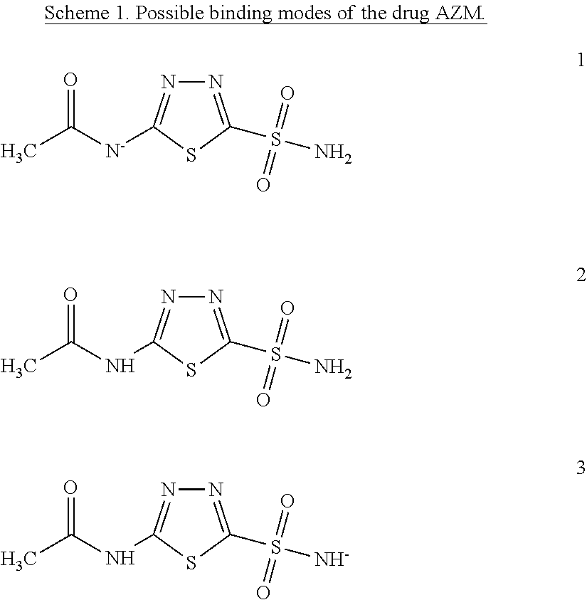
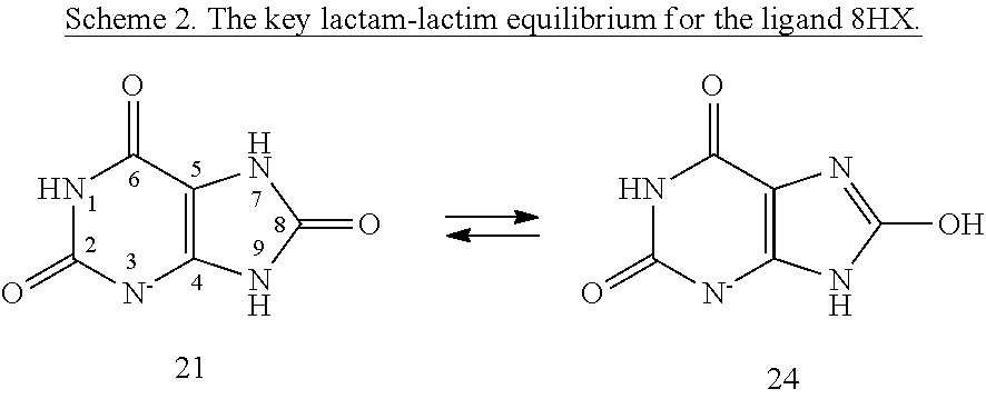
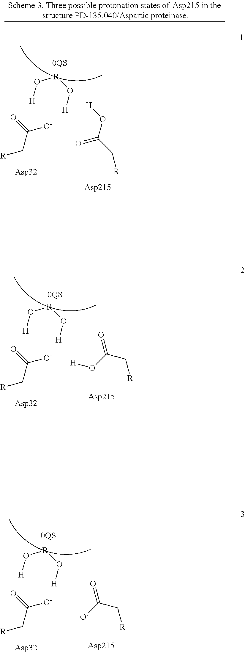
[0008]Within structure-guided drug discovery (SGDD) and structure-based drug discovery (SBDD), accurate understanding of the protein:ligand complex structure, including the relevant proper protonation, is crucial for obtaining meaningful results from docking / scoring, thermodynamic calculations, active site exploration, lead optimization, and ultimately, medicinal chemistry. The most ubiquitous element in the universe is hydrogen, and these protons are critical for exploring the chemistry within the active site. For example, in the drug Mirapex, which is used to treat symptoms of Parkinson's disease, the important chemical activity is conferred by a single aminothiazole tautomeric state rather than an alternative imino tautomer; thus the selection of the wrong state during the drug design would lead to irrelevant findings. This situation is not uncommon, and drug discovery frequently hinges on the determination of one state vs. another state. The present invention provides a verifia...


PUM

PropertyMeasurementUnit
quantum mechanical refinementaaaaaaaaaa
conformationalaaaaaaaaaa
solvent effectaaaaaaaaaa

Abstract

The invention is a diagnostic which overlays quantum mechanical analysis to x-ray crystallography data from one or more proteins to assess and identify the real world conformation, protonation and solvent effects of one or more moieties in said protein. This “overlay” occurs by scoring and identifying the protomer / tautomer states of the moieties using quantum mechanical analysis. The diagnostic results of the present invention accurately identify protein-ligand binding, rendered as an output to a user of a computer in which the x-ray crystallography data is analysed with semi-empirical Hamiltonian quantum mechanics and

Description

CROSS REFERENCE TO RELATED APPLICATIONS[0001]This patent application claims priority to, and incorporates herein by reference, U.S. Provisional Application for Patent No. 62 / 157,787 filed 6 May 2015 and U.S. Provisional Application for Patent No. 62 / 112,951, filed 6 Feb. 2015.STATEMENT OF FEDERALLY SPONSORED RESEARCH[0002]This invention was made in part with government support awarded by the National Institutes of Health, contract number SBIR #R42GM079899. The United States government may have certain rights in and to the invention.BACKGROUND OF THE INVENTION[0003]Field of the Invention[0004]The invention pertains to identifying real-world conformational, protonation and solvent effects of proteins further to identify binding characteristics between one or more proteins and targets therefore, such as drug molecules or other active agents or ligands.[0005]Description of Related Art[0006]Identifying in advance how a drug molecule will interact with conformations of its target protein ...

Claims


Application Information

Patent Timeline
Patent Type & Authority Applications(United States)
IPC IPC(8): G06F19/16G01N23/20G01N33/68G06F19/18G16B15/00G16B20/30
CPCG06F19/16G01N23/20008G01N2223/612G06F19/18G01N2223/304G01N33/6803G16B15/00G16B20/00G16B20/30
Inventor WESTERHOFF, LANCE MICHAELBORBULEVYCH, OLEH Y.MARTIN, ROGER ISAAC
Owner QUANTUMBIO INC
Why Eureka
Try Eureka