Systems providing images guiding surgery

a technology of system and image, applied in the field of providing images for surgery, can solve the problems of difficult to determine the position of a surgical knife and a handheld ultrasound guide during tumour resection, and the difficulty of applying the same handheld ultrasound guidance during tumour resection

Active Publication Date: 2020-01-16
KONINKLJIJKE PHILIPS NV
View PDF0 Cites 1 Cited by
  • Summary
  • Abstract
  • Description
  • Claims
  • Application Information

AI Technical Summary

Benefits of technology

[0031]By means of a computer program product in accordance with an embodiment, real time processing of imaging data and tracking data allow for real time adaptation of visualizations to changes in the region of interest.

Problems solved by technology

Applying the same handheld ultrasound guidance during a tumour resection is much more difficult due to the high deformability of for example a breast and the limited visibility of the tumour boundary.
As a result, it is very difficult to determine the position of a resection tool such as a surgical knife with respect to the tumour boundary.

Method used


Image

  • Systems providing images guiding surgery
  • Systems providing images guiding surgery
  • Systems providing images guiding surgery

Examples


Embodiment Construction

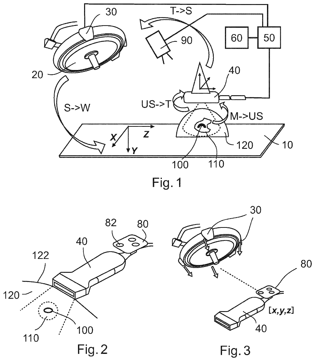
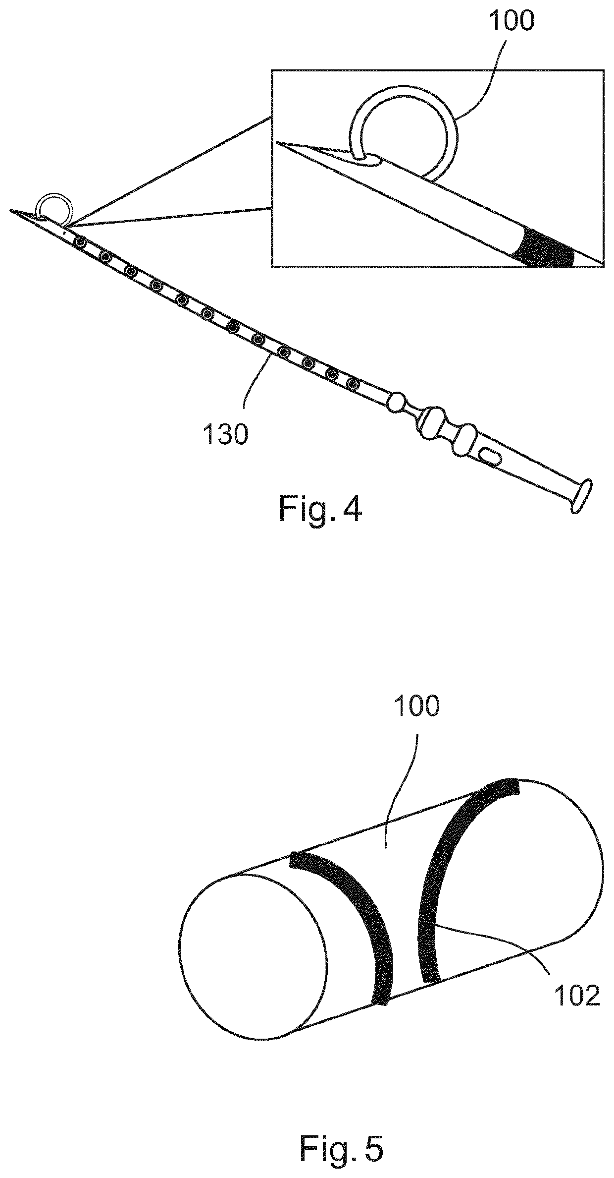
[0047]FIG. 1 illustrates a system in accordance with an embodiment. FIGS. 2 and 3 show elements of the system in more detail. Shown in FIG. 1 is a patient couch or table 10 and an operation light 20, which are typically present in an operation theater. The system according to an embodiment described herein comprises a tracking device 30, an ultrasound device 40, a processing unit 50 and a display 60 as a visualization device. Schematically visualized in FIG. 1 is a marker 100 placed within a region of interest 110 inside a body 120.

[0048]Optionally, the system may comprise at least one camera 90. The camera 90 may be a video camera for imaging the outer surface of the body or may additionally or alternatively be a camera allowing imaging outside the visible spectrum of light. Images generated by any of these cameras may be used to generate an overlay image of the camera image with an ultrasound image from the ultrasound device and / or with a pre-operatively generated image. The posit...


PUM


Abstract

A system may generally comprise a tracking device, an ultrasound device and a processing unit. A position and orientation of the ultrasound device may be traceable by the tracking device. The processing unit may be configured (i) to receive 3D information of a region of interest in relation to a marker, with both the region of interest and the marker being located within a body, (ii) to determine the position of the marker relative to the ultrasound device based on an ultrasound image of the body including the marker, and (iii) to determine the position and orientation of the ultrasound device relative to the tracking device. The system may further comprise a visualization device and the processing unit may further be configured to generate a visualization of the region of interest in relation to an outer surface of the body.

Description

FIELD OF THE INVENTION[0001]The invention generally relates to aspects of assisting and guiding a physician in surgery. Particularly, the invention relates to a system providing visualizations which may help a physician in performing a surgical intervention.BACKGROUND OF THE INVENTION[0002]For example, surgical removal of breast tumours (e.g. lumpectomy) is one of the most frequently performed surgical resections with significant rate increase, due to the leading incidence and prevalence of the breast neoplasms. The clinical goal of the breast surgery is to remove the tumorous tissue entirely without leaving behind more than focally positive margins. Any trace of tumorous tissue left behind would potentially cause tumour recurrence and subsequent revision surgery. Radiotherapy is another treatment option where small microscopic portions left behind are treated by ionizing radiation.[0003]A typical workflow for breast tumour resection starts with the inspection of the 2D images to ma...

Claims


Application Information

Patent Timeline
Patent Type & Authority Applications(United States)
IPC IPC(8): A61B8/00A61B34/20A61B8/08A61B34/10
CPCA61B34/10A61B2034/2063A61B34/20A61B8/4245A61B8/085G06T7/13
Inventor HENDRIKS, BERNARDUS HENDRIKUS WILHELMUSBABIC, DRAZENKOSPLIETHOFF, JARICH WILLEMBYDLON, TORRE MICHELLETOPOREK, GRZEGORZ ANDRZEJPOPOVIC, ALEKSANDRAREICH, CHRISTIAN
Owner KONINKLJIJKE PHILIPS NV
Why Eureka
Try Eureka