
Devices and methods for cellular secretion analysis

Pending Publication Date: 2020-05-21
THE UNIV OF BRITISH COLUMBIA
View PDF2 Cites 3 Cited by
  • Summary
  • Abstract
  • Description
  • Claims
  • Application Information

AI Technical Summary

Benefits of technology

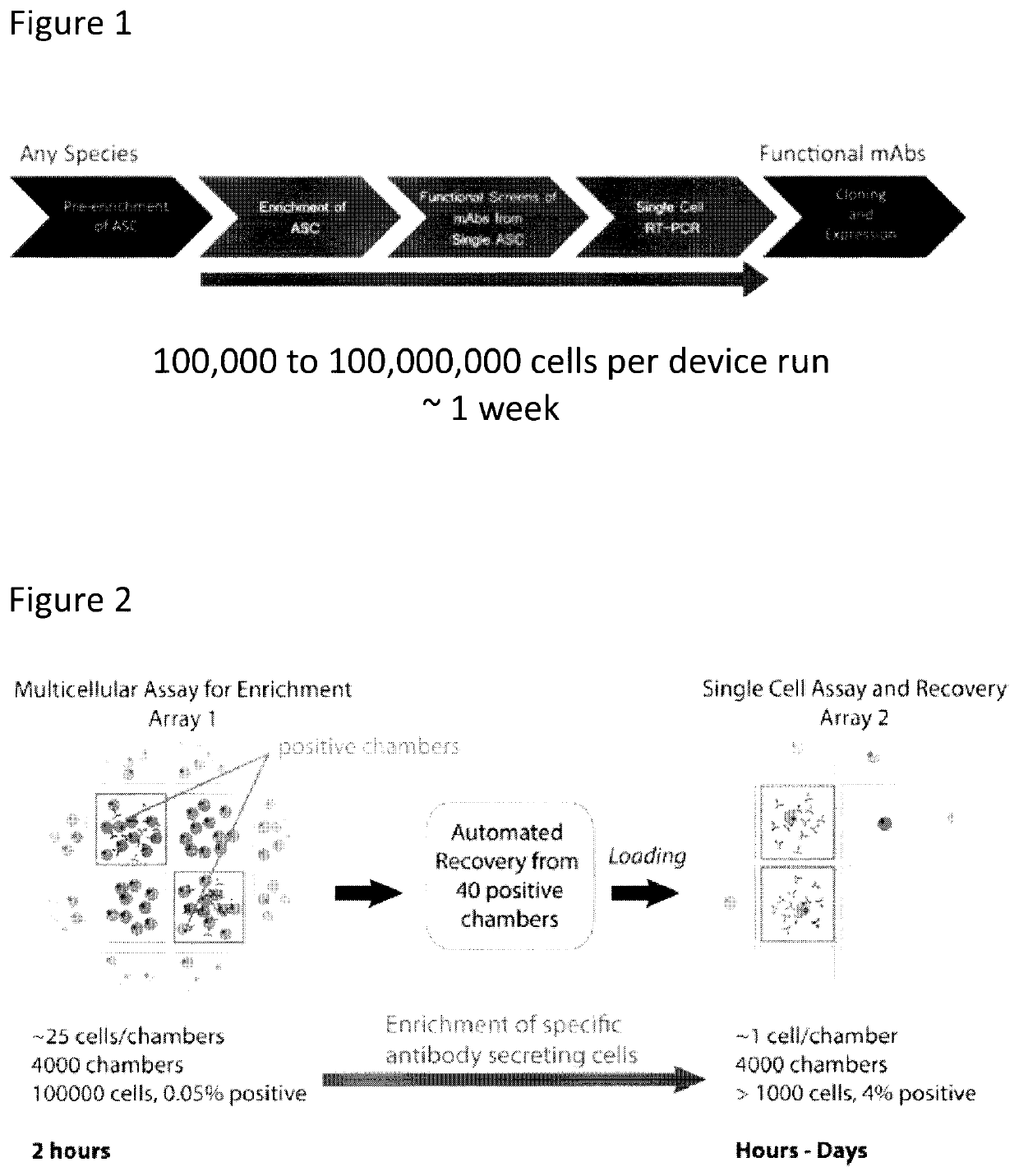
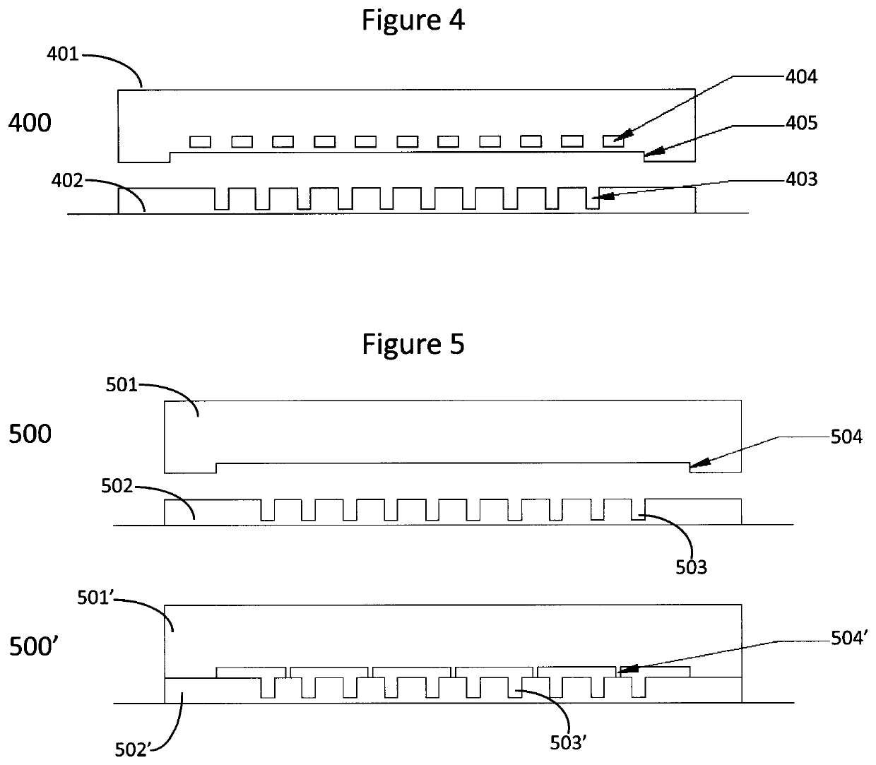
This patent describes a platform for studying the effects of a single cell on other cells. This platform consists of two components: a microfluidic device with open chambers and a sealable connection between them. The device is designed to hold individual cells or small populations of cells. The platform is used to identify which cells are responsible for certain effects, such as cell proliferation or infection. The method involves incubating cells and particles within the open chambers and measuring the effect. The technical effect of this patent is a more efficient and reliable way to analyze the effects of specific cells on other cells.

Problems solved by technology

The discovery of antibodies with optimal therapeutic properties, and in particular, antibodies that target surface receptors, remains a serious bottleneck in drug development.
However, even if an ASC is analyzed individually, and not within a bulk population of cells, because a single ASC generates only a minute amount of antibody, when analyzed in the volume of conventional assay formats, the antibody is too dilute, making it completely undetectable.
Although techniques exist for single ASC analysis, they are still plagued by low throughput, sensitivity and the types of characterizations that can be carried out.

Method used


Image

  • Devices and methods for cellular secretion analysis
  • Devices and methods for cellular secretion analysis
  • Devices and methods for cellular secretion analysis

Examples


example 1

oading into Two-Component Microfluidic Device

[0305]The efficiency of loading a two-component microfluidic device comprising arrays of individual microfluidic chambers was assessed by loading multiple types of microparticles into the open chambers of the bottom component of the device. The dimensions of the microfluidic chambers were 80 μm×120 μm×160 μm (width, length, height).

[0306]Five populations of microparticles, each consisting of 5 μm diameter polystyrene beads labeled with a different concentration of fluorophore, were prepared at even concentrations of approximately 1,000,000 beads per mL. Each of the five populations were mixed together in a single tube at equal volumetric ratios. A 100 μL aliquot of the mixture containing approximately 100,000 total microbeads was introduced over a section of the bottom layer of the two-component microfluidic device having approximately 10,000 total chambers. This corresponded to approximately 10 beads per chamber.

[0307]The device was then...

example 2

ing into Two-Component Microfluidic Devices

[0314]The loading of adherent readout cells was assessed in a two-component device of the invention, and compared to the loading of the same cells into a preassembled microfluidic device fabricated via MSL.

[0315]The loading of an adherent cell line (Tango™ CXCR4-bla U2OS) into pre-assembled microfluidic device was assessed by introducing cells through an inlet port and flowing them into the individual chambers through microfluidic channels (channel width 100 μm). This resulted in poor chamber loading uniformity with increased concentrations of cells in the center of the device, low cell loading in the corners of the device, and large variability in the cell loading density between adjacent chambers. Channel clogging due to cell sticking was observed even in medium conditions that included trypsin (FIG. 32). Further, once loaded into chambers and after incubation, cells did not plate down well and showed morphology consistent with stress. Ce...

example 3

Chamber Aspect Ratio During Medium Exchange

[0318]A series of experiments were conducted to evaluate the influence of chamber aspect ratio on two-component microfluidic device performance during the exchange of chamber medium. In one experiment, microfluidic devices having chambers with differing aspect ratios (ratio of the depth / height to the minimum lateral dimension), were tested to determine if medium exchange by providing solution over chambers containing cells or beads results in loss of the cells or beads from chambers, or displacement of the cells or beads within chambers.

[0319]A device having chambers with dimensions of 100 μm×100 μm×150 μm (depth / height×width×length), corresponding to an aspect ratio of about 1, and channel dimensions of 20 μm×100 μm (height×width) were tested as differing flow rates. Fluid ports at the inlet and the outlet of the device were connected to pressure regulators and the pressure was adjusted to modulate the flow rate through the channels. It wa...


PUM


Abstract

Methods and devices for identifying a cell population comprising an effector cell exhibiting an extracellular effect are provided. The method comprises retaining in a plurality of open chambers a plurality of cell populations, each optionally comprising one or more effector cells. The open chambers can each comprise a readout particle population, and the open chambers are present in a first component of a device comprising a first component and optionally a second component. The open chambers have an average aspect ratio of ≥0.6 and the first component forms a reversible seal with the second component. The method further comprises incubating the plurality of cell populations or a subset thereof, and the one or more readout particles, or a subset thereof, within the chambers, assaying the cell populations for the presence of the extracellular effect, wherein the readout particle(s) provides a direct or indirect readout of the extracellular effect, and determining, based on the results of the assaying step, whether one or more cells within one or more cell populations of the plurality exhibits the extracellular effect.

Description

CROSS-REFERENCE TO RELATED APPLICATION[0001]This application claims priority from U.S. Provisional Patent Application Ser. No. 62 / 309,663, filed Mar. 17, 2016, the disclosure of which is incorporated by reference in its entirety.BACKGROUND OF THE INVENTION[0002]The cell is the fundamental unit of life and no two cells are identical. Indeed, seemingly identical clonal populations of cells have been shown to display phenotypic differences among cells within the population. Cellular differences exist across all levels of life, ranging from bacterial cells to partially differentiated cells (for example, adult stem and progenitor cells) to highly differentiated mammalian cells (for example, immune cells such as antibody secreting cells (ASCs)). Differences in cellular state, function and responses can arise from a variety of mechanisms including different histories, different differentiation states, epigenetic variations, cell cycle effects, stochastic variations, differences in genomic ...

Claims


Application Information

Patent Timeline
IPC IPC(8): B01L3/00G01N33/543C12Q1/04
CPCB01L2400/06B01L3/502707C12Q1/04B01L2300/0809B01L2400/0403G01N33/54366B01L3/502761B01L2200/0647B01L2200/12B01L3/50273G01N33/56966G01N33/6854B01L2300/0819C12Q1/00C12Q1/02G01N33/483C12M23/16C12M23/34C12M41/46G01N2015/1006G01N15/1433G01N33/5005G01N33/6872
Inventor HANSEN, CARL LARS GENGHISLISAINGO, KATHLEENLECAULT, VERONIQUE
Owner THE UNIV OF BRITISH COLUMBIA
Learn More