A laparoscopic adapter, an echocardiography probe and a method for coupling the adapter to the probe

a technology of echocardiography and adapter, which is applied in the field of laparoscopic procedures, can solve the problems of lack of volumetric information, surgeons losing tactile feedback, orientation and all other information normally provided during conventional surgery, and achieve the effect of facilitating distinction

Inactive Publication Date: 2020-06-18
KONINKLJIJKE PHILIPS NV
View PDF0 Cites 2 Cited by
  • Summary
  • Abstract
  • Description
  • Claims
  • Application Information

AI Technical Summary

Benefits of technology

[0017](13) the terms “signal”, “data” and “command” broadly encompasses all forms of a detectable physical quantity or impulse (e.g., voltage, current, or magnetic field strength) as understood in the art of the present disclosure and as exemplary described in the present disclosure for transmitting information and/or instructions in support of applying various inventive principles of the present disclosure as subsequently described in the present disclosure. Signal/data/command communication between various components of the present disclosure may involve a

Problems solved by technology

During minimally invasive laparoscopic procedures, any endoscopic feedback provided to a surgeon has proved to be limited.
Therefore surgeons lose their tactile feedback, sense of orientation and all other information that are usually provided during conventional surgery.
A clear disadvantage of 2D ultrasound is lack of volumetric information which could be of interest for precise assessment of the region of interest of the o

Method used


Image

  • A laparoscopic adapter, an echocardiography probe and a method for coupling the adapter to the probe
  • A laparoscopic adapter, an echocardiography probe and a method for coupling the adapter to the probe
  • A laparoscopic adapter, an echocardiography probe and a method for coupling the adapter to the probe

Examples


second embodiment

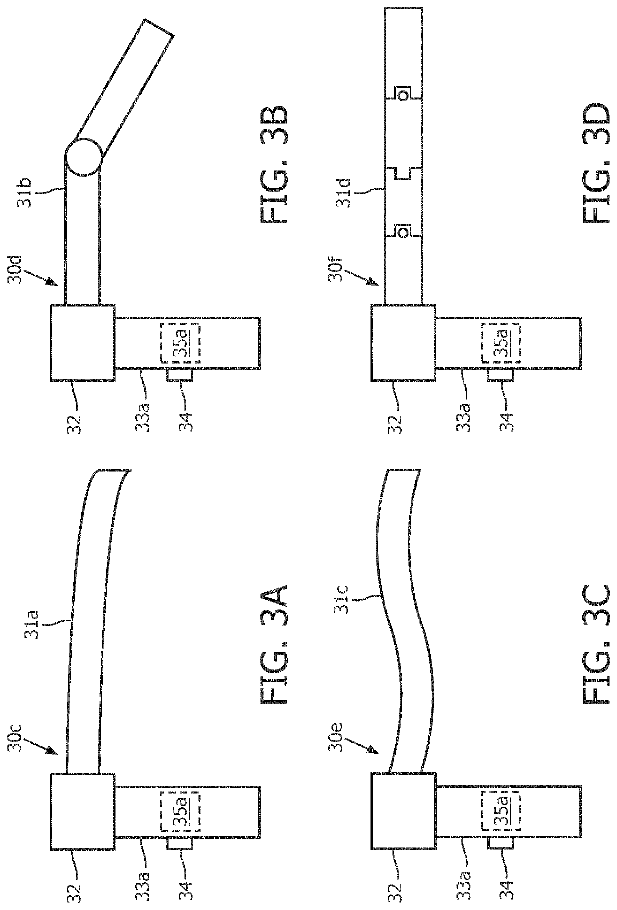
[0052]In a second embodiment, laparoscopic sleeve 31 may have a rigid material composition with a jointed cylindrical configuration 31b as shown in FIG. 3B for facilitating a manual pivoting of a distal end of laparoscopic sleeve 31.

third embodiment

[0053]In a third embodiment, laparoscopic sleeve 31 may have a semi-rigid material composition with a cylindrical configuration, such as, for example, laparoscopic sleeve 31 may be a catheter 31c as shown in FIG. 3C.

fourth embodiment

[0054]In a fourth embodiment, laparoscopic sleeve 31 may have a rigid material composition with a controllable cylindrical configuration, such as, for example, laparoscopic sleeve 31 may be a snake robot 31d as shown in FIG. 3D.

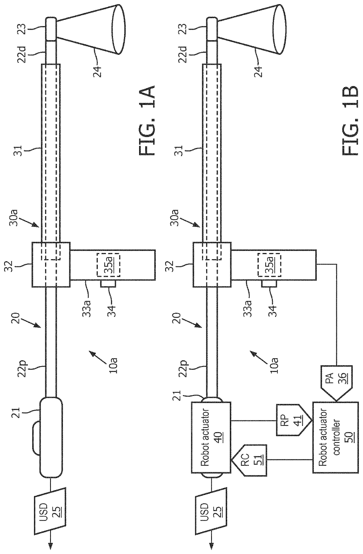
[0055]Referring back to FIG. 1A, adapter mount 32 has a mount channel for receiving flexible shaft 20 and / or laparoscopic sleeve 31 as symbolically shown by the dashed lines within laparoscopic sleeve 31.

[0056]In practice, adapter mount 32 may clamp onto flexible shaft 20 and / or laparoscopic sleeve 31 to maintain laparoscopic sleeve 31 at a fixed position relative to ultrasound transducer 23.

[0057]Still referring to FIG. 1A, probe handle 33a is affixed to adapter mount 32 to enable a user manipulation of a coarse alignment of ultrasound transducer 23 to a region of interest of a patient anatomy.

[0058]Probe handle 33a includes a user input device 34 (e.g., a joystick, a rollerball, etc.) for operating a probe controller 35a to generate a probe actuation signal...


PUM


Abstract

A 3D echocardiography probe (20) (e.g., a 3D transesophageal echocardiography probe or a 3D intracardiac echocardiography probe) can be adapted into a 3D laparoscopic ultrasound probe (10a) for laparoscopic procedures. The 3D echocardiography probe includes a flexible shaft (22d, 22p) to which a laparoscopic adapter (30a) can be coupled. The laparoscopic adapter comprises a laparoscopic sleeve (31) configured to partially encircle a portion of the flexible shaft of the 3D echocardiography probe and a probe handle (33a) mountable to the laparoscopic sleeve (31).

Description

FIELD OF THE INVENTION[0001]The present disclosure generally relates to laparoscopic procedures (e.g., prostatectomy, aplenectomy, nephrectomy and hepatectomy). The present disclosure specifically relates to an adaption of a three-dimensional (“3D”) echocardiography probe into a 3D laparoscopic ultrasound (“LUS”) probe for the performance of a laparoscopic procedure.BACKGROUND OF THE INVENTION[0002]During minimally invasive laparoscopic procedures, any endoscopic feedback provided to a surgeon has proved to be limited. Specifically, the surgeon using endoscopic camera may only view an outer surface of an organ. Therefore surgeons lose their tactile feedback, sense of orientation and all other information that are usually provided during conventional surgery. To overcome this endoscopic limitation, two-dimensional (“2D”) laparoscopic ultrasound (“LUS”) probes designed to have direct contact with a parenchyma of an organ were introduced to provide an intra-operative visualization of t...

Claims


Application Information

Patent Timeline
IPC IPC(8): A61B8/12A61B8/00A61B8/08A61B46/10
CPCA61B8/467A61B2034/301A61B1/0014A61B1/008A61B8/4466A61B46/10A61B1/00135A61B8/483A61B8/4254A61B8/0883A61B8/445A61B1/3132A61B8/12A61B1/00147A61B8/4218A61B8/4411A61B8/54
Inventor TOPOREK, GRZEGORZ ANDRZEJPOPOVIC, ALEKSANDRA
Owner KONINKLJIJKE PHILIPS NV
Why Eureka
Try Eureka