A system and method for automated labeling and annotating unstructured medical datasets

a medical dataset and automated labeling technology, applied in healthcare informatics, instruments, computing models, etc., can solve the problems of increasing the demand for diagnostic imaging, increasing the risk of human error and delayed diagnosis, and remaining limited, and the labeling requirement is inherently difficult to m

Active Publication Date: 2020-09-10
THE GENERAL HOSPITAL CORP
View PDF3 Cites 6 Cited by
  • Summary
  • Abstract
  • Description
  • Claims
  • Application Information

AI Technical Summary

Benefits of technology

[0008]The present disclosure addresses the aforementioned drawbacks by providing a system and method for using supervised and unsupervised learning schemes to automatically label medical images for use in subsequent deep learning applications. The system can generate a large labeled dataset from a small initial training set using an iterative snowball sampling scheme. A machine-learning powered, automatic organ classifier for imaging datasets, such as CT datasets, with a deep convolutional neural network (CNN) followed by an organ dose calculation is also provided. This technique can be used for patient-specific organ dose estimation because the locations and sizes of organs for each patient can be calculated independently, rather than other simulation based methods.

Problems solved by technology

The increased demand for diagnostic imaging also presents a growing risk for human error and delayed diagnosis.
While computer aided detection (CADe) and diagnosis (CADx) systems can reduce such problems, they remain limited due to their reliance upon hand-crafted features.
This labeling requirement is inherently difficult to meet in the medical domain where medical expertise is expensive, labeling is tedious and time-consuming, and examples of certain disease pathologies may be rare.
These approaches are limited because only low-level visual features such as color, edges, and color layouts are extracted.
Even with higher-level feature extraction from MR voxels by hierarchical learning using two-layer random forests, segmentation performance is not generally better than features extracted by deep convolutional neural networks.
Furthermore, all current methods of annotating medical images still require mid- to large-sized labeled image datasets for obtaining the trained model.
Classifying the location is a challenging problem because the details of body regions can vary dramatically between patients, such as with brain gyral patterns, cervical vertebral anatomy, pulmonary vessels, and bowel distribution.
Degenerative changes can also distort bony anatomy enough to confuse the network.
However, creating high-quality training datasets is expensive and time-consuming.

Method used


Image

  • A system and method for automated labeling and annotating unstructured medical datasets
  • A system and method for automated labeling and annotating unstructured medical datasets
  • A system and method for automated labeling and annotating unstructured medical datasets

Examples


Embodiment Construction

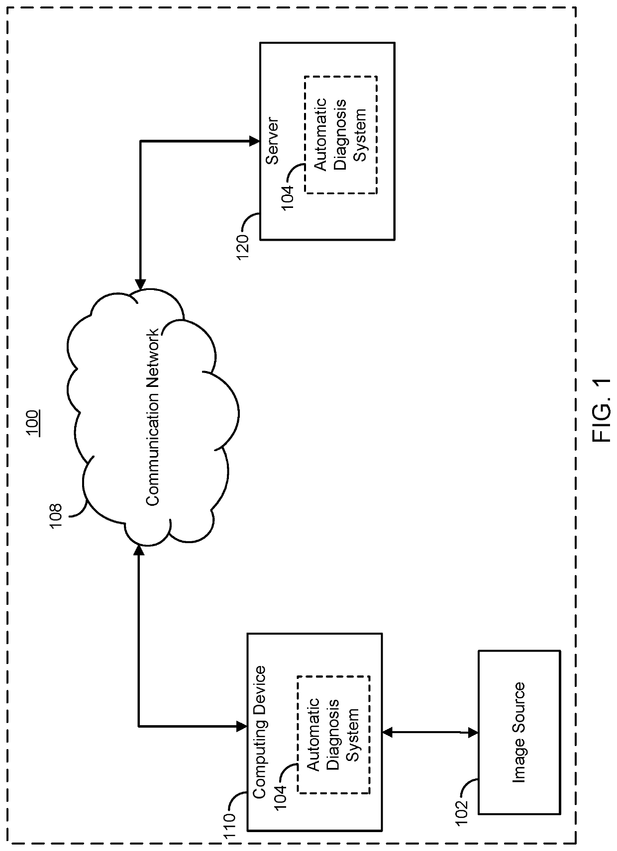
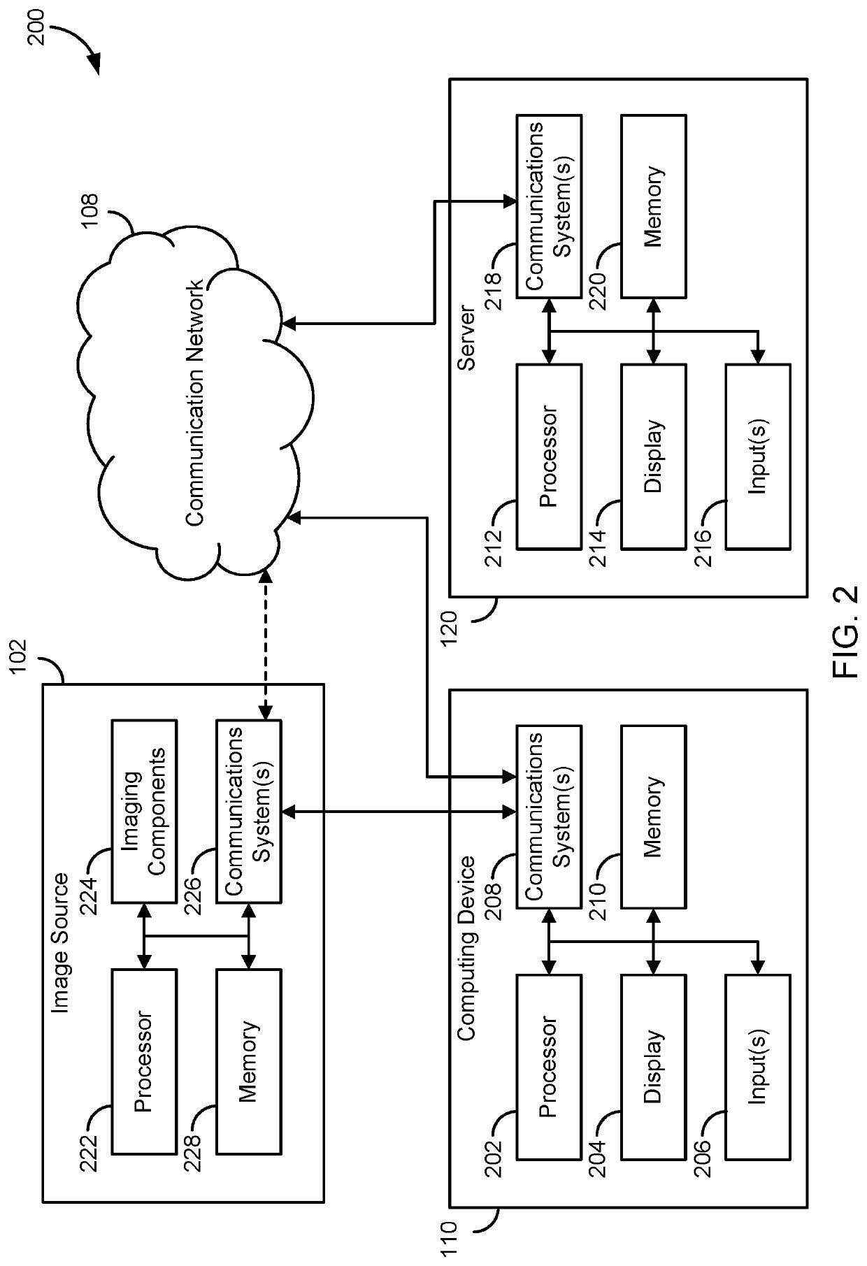
[0028]The present disclosure provides systems and method for supervised and unsupervised learning schemes that may be used to automatically label medical images for use in deep learning applications. Large labeled datasets may be generated from a small initial training set using an iterative snowball sampling scheme. A machine learning powered automatic organ classifier for imaging datasets, such as CT datasets, with a deep convolutional neural network (CNN) followed by an organ dose calculation is also provided. This technique can be used for patient-specific organ dose estimation since the locations and sizes of organs for each patient can be calculated independently.

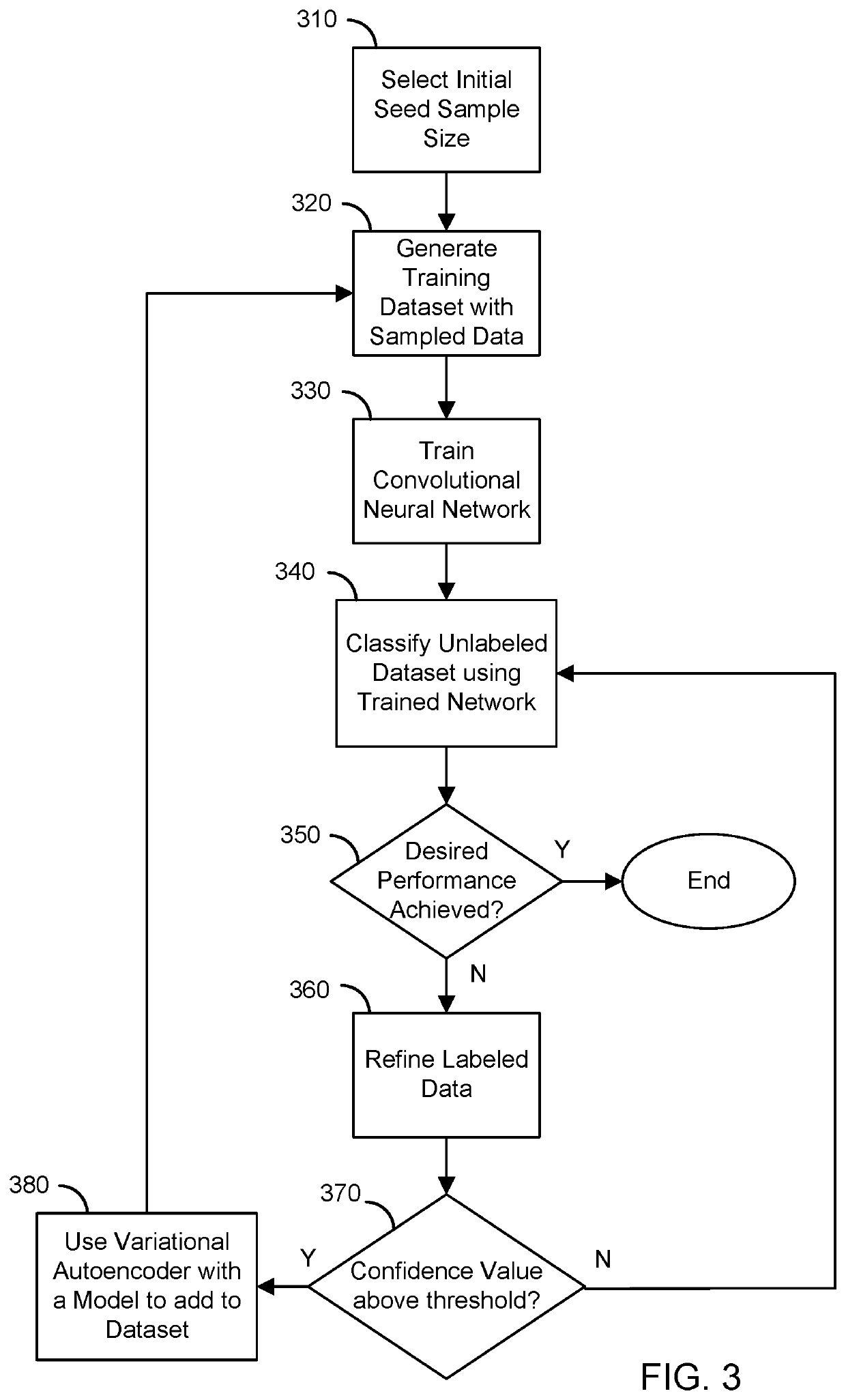
[0029]In one configuration, a desired classification accuracy may be achieved with a minimal labeling process. Using an iterative snowball sampling approach, a large medical image dataset may be annotated automatically with a smaller training subset. The automatic labeling system may include a variational autoencoder ...


PUM


Abstract

Supervised and unsupervised learning schemes may be used to automatically label medical images for use in deep learning applications. Large labeled datasets may be generated from a small initial training set using an iterative snowball sampling scheme. A machine learning powered automatic organ classifier for imaging datasets, such as CT datasets, with a deep convolutional neural network (CNN) followed by an organ dose calculation is also provided. This technique can be used for patient-specific organ dose estimation since the locations and sizes of organs for each patient can be calculated independently.

Description

CROSS-REFERENCE TO RELATED APPLICATIONS[0001]This application claims the benefit of U.S. Provisional Patent Application Ser. No. 62 / 555,799 filed on Sep. 8, 2017, and entitled “A methodology for automated labeling and annotation for unstructured big medical datasets,” and U.S. Provisional Patent Application Ser. No. 62 / 555,767 filed on Sep. 8, 2017, and entitled “Method and apparatus of machine learning based personalized organ dose estimation.STATEMENT REGARDING FEDERALLY SPONSORED RESEARCH[0002]N / ABACKGROUND[0003]Diagnostic medical imaging has become central to the practice of modern medicine and diagnostic examination volume has increased during the past decade. Additionally, as systems have become more advanced with higher resolution, the number of images in a given study has also increased. The increased demand for diagnostic imaging also presents a growing risk for human error and delayed diagnosis. While computer aided detection (CADe) and diagnosis (CADx) systems can reduce ...

Claims


Application Information

Patent Timeline
Patent Type & Authority Applications(United States)
IPC IPC(8): G06K9/62G06K9/52G16H30/40G16H15/00G16H20/40G06V10/764
CPCG06K9/52G16H15/00G16H30/40G06K2209/051G16H20/40G06K9/6262G06K9/6257G06V10/454G06V2201/031G06V10/82G06V10/764G06N3/084G06V20/20G06F18/217G06F18/2148G06F18/2155G06F18/2415G06F18/2431G06N7/01G06V10/42G06V10/751
Inventor DO, SYNHOCHO, JUNG HWAN
Owner THE GENERAL HOSPITAL CORP
Why Eureka
Try Eureka