
Analysis method for breast image and electronic apparatus using the same

an analysis method and breast image technology, applied in image enhancement, instruments, ultrasonic/sonic/infrasonic image/data processing, etc., can solve the problems of time-consuming and ineffective examination for medical personnel

Pending Publication Date: 2021-11-04
TAIHAO MEDICAL
View PDF8 Cites 1 Cited by
  • Summary
  • Abstract
  • Description
  • Claims
  • Application Information

AI Technical Summary

Benefits of technology

The patent provides an analysis method and electronic apparatus for breast images that can detect and analyze abnormal regions of interest quickly and effectively. The method uses a detection model based on rectangular features of breast images to identify the abnormal region and display related property analysis. This allows for the identification of abnormal symptoms with a single analysis method, even when dealing with a large number of breast ultrasound images.

Problems solved by technology

However, when there are a massive number of the breast images, it is very time-consuming and ineffective for medical personnel to examine whether the aberrant part is included in the breast images one by one.
On the other hand, mistakes can often be made if the aberrant part in the breast image is filtered only by the human eye.

Method used


Image

  • Analysis method for breast image and electronic apparatus using the same
  • Analysis method for breast image and electronic apparatus using the same
  • Analysis method for breast image and electronic apparatus using the same

Examples


Embodiment Construction

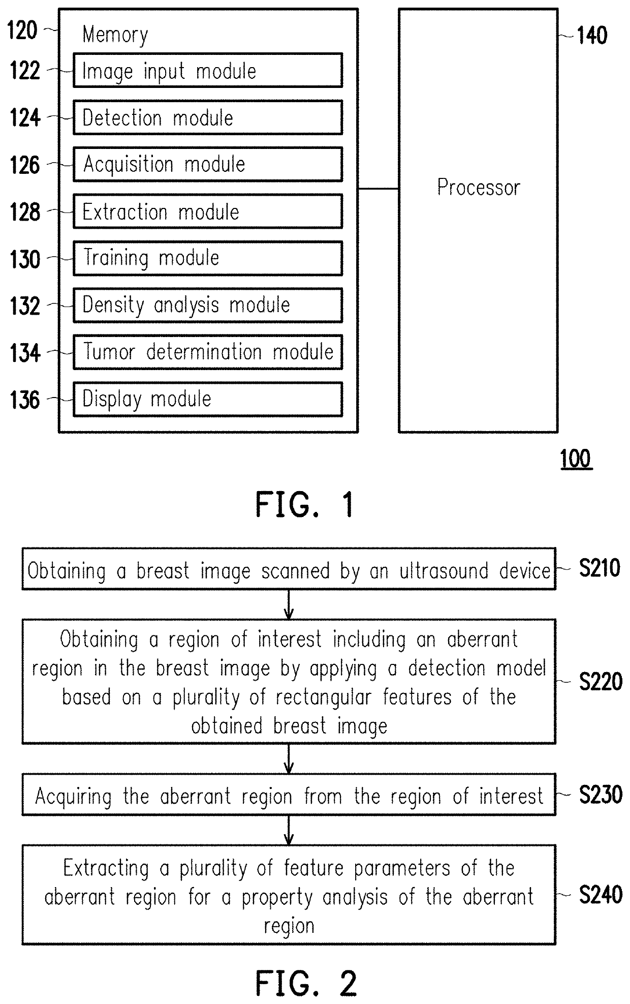
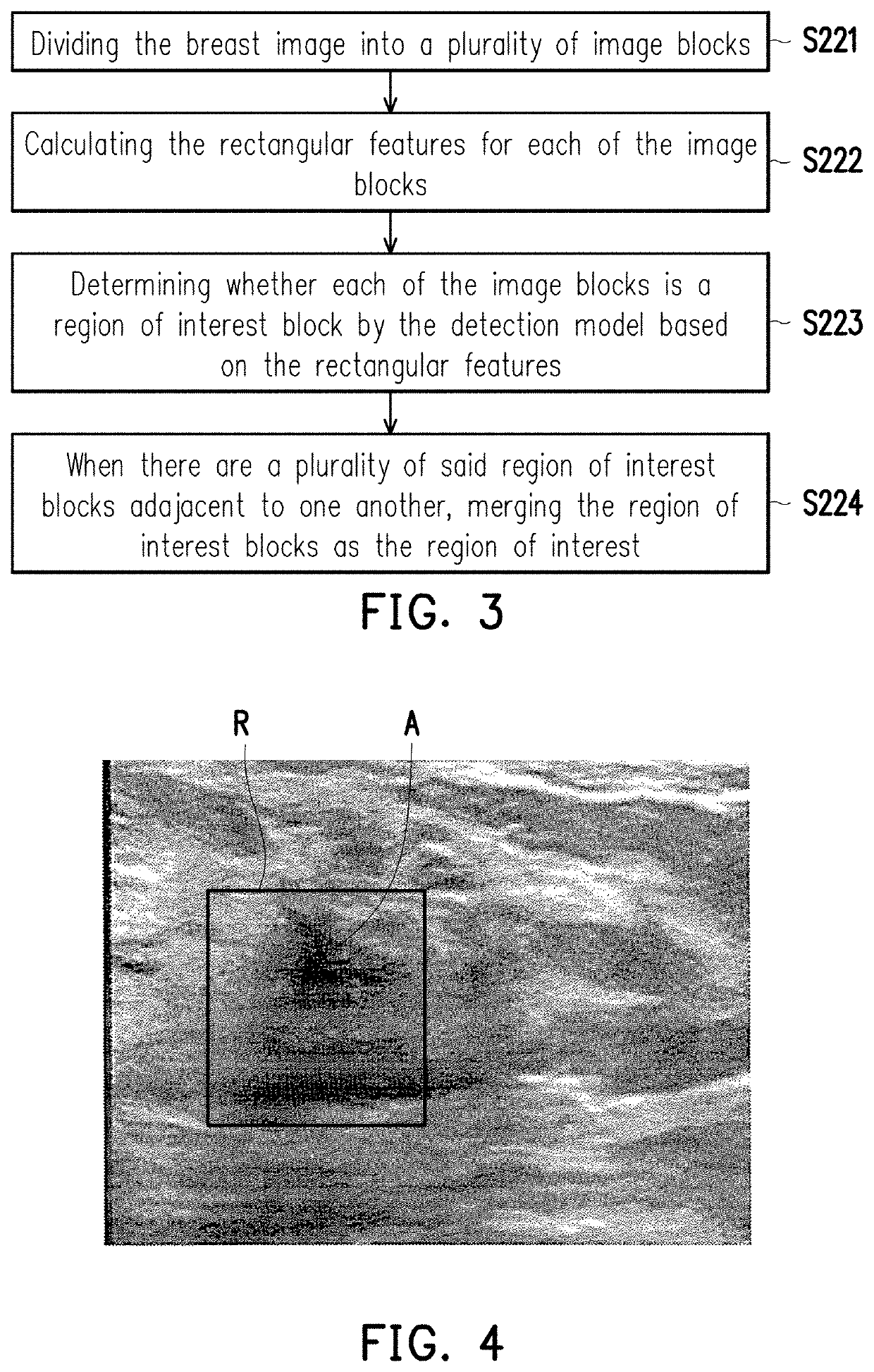
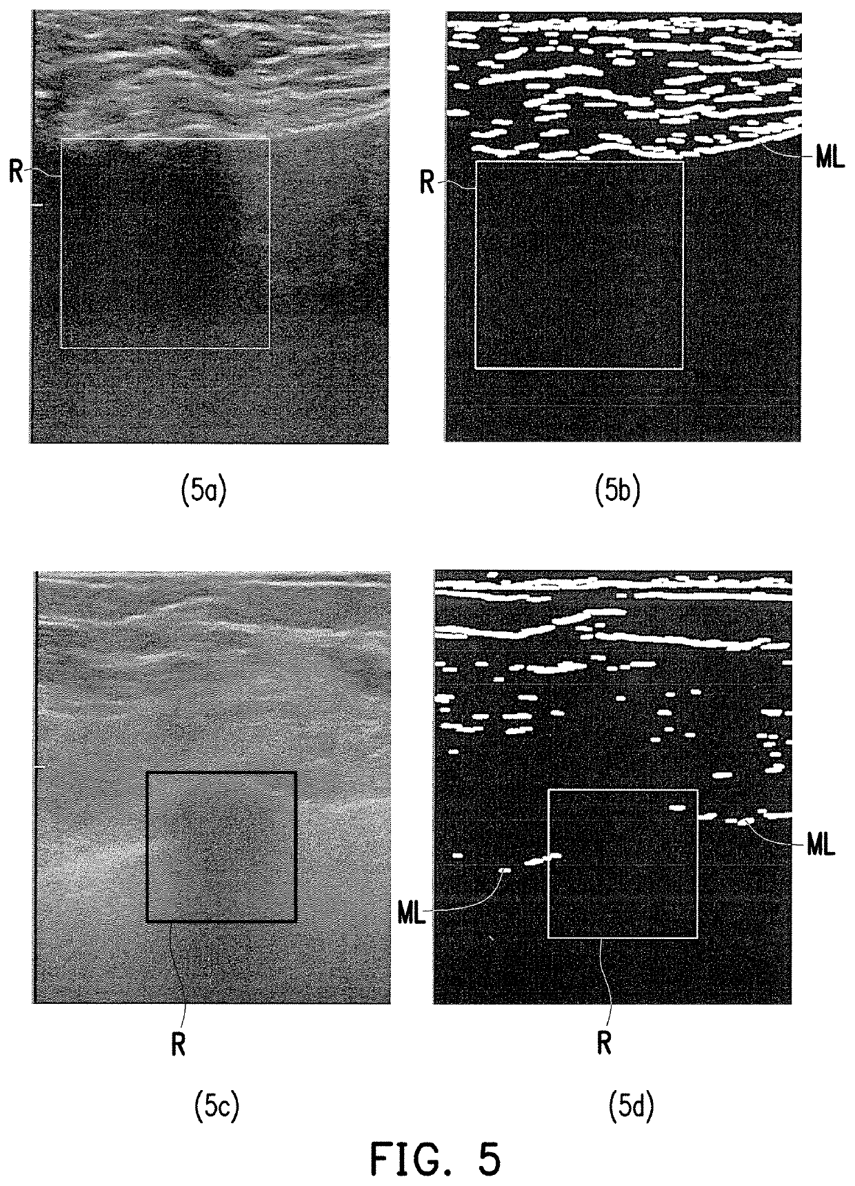
[0031]Reference will now be made in detail to the present preferred embodiments of the invention, examples of which are illustrated in the accompanying drawings. Wherever possible, the same reference numbers are used in the drawings and the description to refer to the same or like parts.

[0032]Some embodiments of the invention are described in details below by reference with the accompanying drawings, and as for reference numbers cited in the following description, the same reference numbers in difference drawings are referring to the same or like parts. The embodiments are merely a part of the invention rather than disclosing all possible embodiments of the invention. More specifically, these embodiments are simply examples of devices and methods recited in claims of the invention.

[0033]In the analysis method for breast image and the electronic apparatus using the same as proposed in the embodiments of the invention, first of all, a region of interest (ROI) including an aberrant reg...


PUM


Abstract

An analysis method and an electronic apparatus for breast image are provided. The method includes the following steps. One or more breast ultrasound images are obtained. The breast ultrasound images are used for forming a three-dimensional (3D) breast model. A volume of interest (VOI) in the breast ultrasound image is obtained by applying a detection model on the 3D breast model. The VOI is compared with a tissue segmentation result. The VOI is determined as a false positive according to a compared result between the VOI and the tissue segmentation result. The compared result includes that the VOI is located at a glandular tissue based on the tissue segmentation result. In response to the VOI being located in the glandular tissue of the tissue segmentation result, the VOI is compared with the lactiferous duct in the 3D breast model.

Description

CROSS REFERENCE TO RELATED APPLICATION[0001]This application is a continuation-in-part application of and claims the priority benefit of U.S. application Ser. No. 15 / 622,077, filed on Jun. 14, 2017, now pending, which claims the priority benefit of Taiwan application serial no. 105120124, filed on Jun. 27, 2016. The entirety of each of the above-mentioned patent applications is hereby incorporated by reference herein and made a part of specification.BACKGROUND OF THE INVENTION1. Field of the Invention[0002]The invention relates to an analysis method and an electronic apparatus using the same, and more particularly, to an analysis method for analyzing breast image and an electronic apparatus using the same.2. Description of Related Art[0003]Mammary carcinoma is a common female malignant tumor, and the main symptom includes breast tumors, abnormal secretions, or shape variation, etc. Screening an aberrant symptom for breast in advance can facilitate in treating the tumor earlier so th...

Claims


Application Information

Patent Timeline
IPC IPC(8): A61B8/08G06T7/11G06T7/149G06T17/00G06V10/143G06V10/25G06V10/50G06V10/75
CPCA61B8/085A61B8/0825A61B8/483G06T7/11G06T2207/30068G06T17/00G06T2207/10132G06T2207/20081G06T7/149A61B8/5223G16H50/30G06V10/143G06V10/25G06V10/50G06V2201/032G16H30/40G16H50/20G16H20/40G16H40/60G06T7/0012G06T2207/10136G06T2207/20084G06V10/75G06F18/22
Inventor HSU, JEN-FENGCHEN, HONG-HAOCHEN, RONG-TAILAI, HSIN-HUNGTENG, WEI-HAN
Owner TAIHAO MEDICAL
Learn More