Method and system to enhance robust identification of abnormal regions in radiographs

a radiograph and abnormal anatomical technology, applied in image analysis, instruments, computing, etc., can solve the problems of premature death in women, visual reading of mammograms, and the possibility of missing breast cancer in its early stages

Inactive Publication Date: 2000-05-23
PITTSBURGH UNIV OF
View PDF16 Cites 76 Cited by
  • Summary
  • Abstract
  • Description
  • Claims
  • Application Information

AI Technical Summary

Problems solved by technology

For example, detection and diagnosis of abnormal anatomical regions in radiographs, such as masses and microcalcifications in womens' breast radiographs (mammograms), are among the most important and difficult tasks performed by radiologists.
Breast cancer is a leading cause of premature death in women over forty years old.
Unfortunately, visual reading of mammograms has two major disadvantages.
First, it is often possible to miss the breast cancer in its early stages.
This is because, unlike many other cancers, there is as yet no clear way to detect premalignant changes in the breast.
This results partly from the relative inaccessibility of breast tissue.
A second disadvantage of visual reading of mammograms is that these readings are both labor intensive, time consuming and subjective.
The use of features from only one level to analyze a growth area, especially from the cross-over level, yields unsatisfactory results since this layer contains more information on background or surrounding regions than on the growth area itself.
At this stage the criteria for finding suspicious regions are not generally strict, therefore these found suspicious regions may contain many false-positive regions.

Method used


Image

  • Method and system to enhance robust identification of abnormal regions in radiographs
  • Method and system to enhance robust identification of abnormal regions in radiographs
  • Method and system to enhance robust identification of abnormal regions in radiographs

Examples


Embodiment Construction

The above processing can be demonstrated with reference to FIGS. 9(a)-9(c), wherein FIG. 9(a) represents a digital radiograph of a human breast (a mammogram). This image is analyzed as follows:

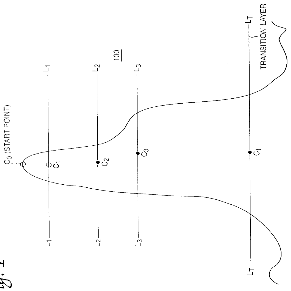
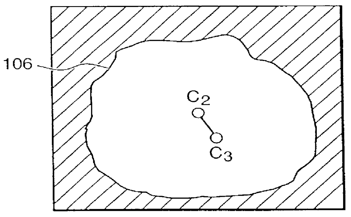
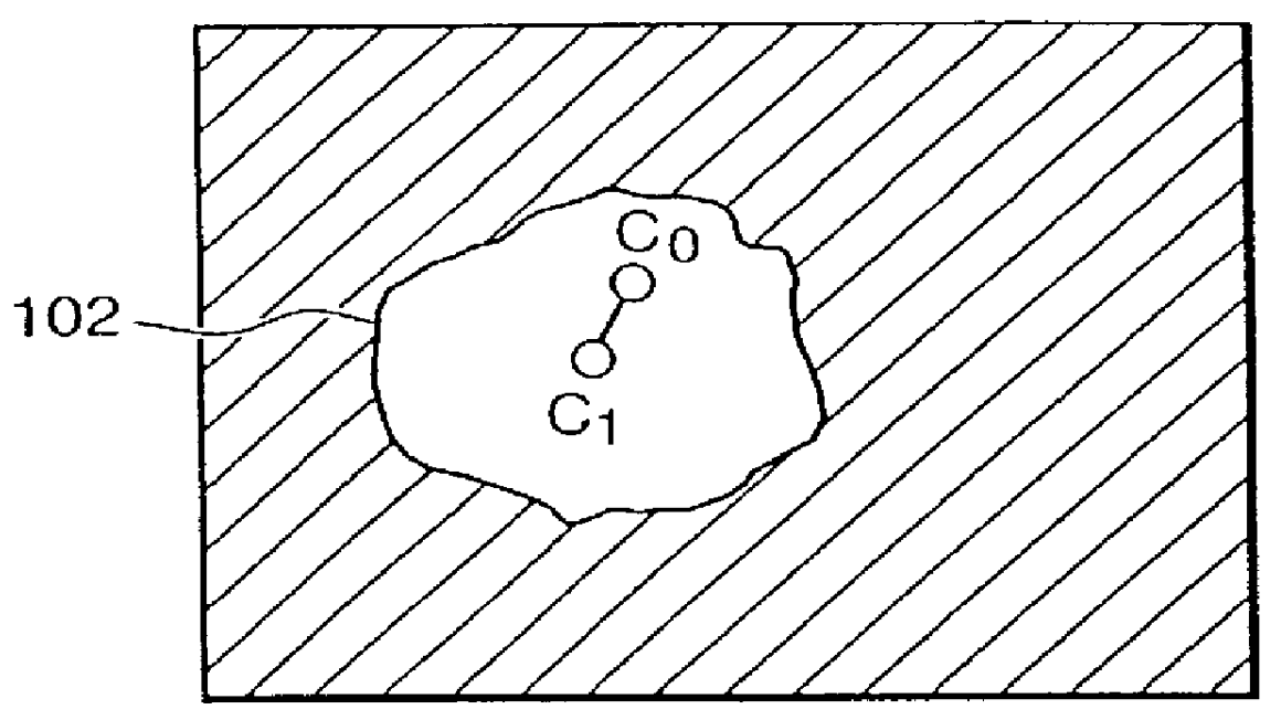
First the image is obtained (step S600) and all suspicious regions (R.sub.1 -R.sub.5) in the image are located (step S610). Then, for each suspicious region, i.e., for each of regions R.sub.1 to R.sub.5, a start point is found (step S640). For example, for suspicious region R.sub.1, the start point P.sub.R1 is found (step S640), following which the various layers for the region R.sub.1 are extracted (step S650). Region R.sub.1 is shown in cross-section in FIG. 9(b) and the first layer (corresponding to line L.sub.1 --L.sub.1 in FIG. 9(b)) is shown in FIG. 9(c).

For each layer the features for that layer are determined. Then the rules are applied to the features in order to determine whether or not the suspicious region is a true-positive region (step S670).

Increased Robustness

In order to increa...


PUM


Abstract

A method and apparatus for detecting abnormal regions in living tissue depicted in a digital radiograph includes identifying suspected abnormal regions depicted in the radiograph and then, for each identified suspected abnormal region, extracting multiple topographic layers of the region from the digital radiograph; determining features of the region in each of the layers; and applying inter-layer multivariate non-linear criteria to the features to determine whether the suspected abnormal region is to be classified as an abnormal region. The distribution of digital values in the digitized radiograph is modified and a probabalistic determination is made as to whether suspected abnormal regions are actually abnormal from multiple analyses. An image is processed using at least two partially correlated detection schemes to produce at least two corresponding result sets of potentially abnormal regions; and the results of the detection schemes are combined using logical "AND" or logical "OR" combining rules to detect abnormal regions in the radiograph.

Description

1. FIELD OF THE INVENTIONThis invention relates to detection of abnormal anatomical regions depicted in radiographs.2. BACKGROUNDDetection and analysis of target objects in digital images are useful and important tasks. For example, detection and diagnosis of abnormal anatomical regions in radiographs, such as masses and microcalcifications in womens' breast radiographs (mammograms), are among the most important and difficult tasks performed by radiologists.Breast cancer is a leading cause of premature death in women over forty years old. Evidence shows that early detection, diagnosis and treatment of breast cancer significantly improves the chances of survival, reducing breast cancer morbidity and mortality. Many methods for early detection of breast cancer have been studied and tested, among them mammography. To date mammography has proven to be the most cost effective means of providing useful information to diagnosticians regarding abnormal features in the breast and potential r...

Claims


Application Information

Patent Timeline
Patent Type & Authority Patents(United States)
IPC IPC(8): G06T7/00
CPCG06T7/0012
Inventor GUR, DAVIDZHENG, BINCHANG, YUAN-HSIANG
Owner PITTSBURGH UNIV OF
Why Eureka
Try Eureka