Method and apparatus for obtaining an electrocardiograph

a technology of electrocardiograph and electrocardiograph plate, which is applied in the field of electrocardiograph method and apparatus, can solve the problems of noisy ecg, difficulty increase, and inability to obtain clear signals

Inactive Publication Date: 2005-03-29
SCHILLER MEDICAL
View PDF4 Cites 34 Cited by
  • Summary
  • Abstract
  • Description
  • Claims
  • Application Information

AI Technical Summary

Benefits of technology

The invention will be more readily understood with reference to the following description of some preferred embodiments cited as non-limitative examples, and explained in conjunction with the attached schematic drawings, wherein:

Problems solved by technology

In capturing physiological signals, obtaining a clear signal often poses a problem, as the signal is generally weak and drowned by background noise.
This difficulty increases when the signal is acquired in an MRI environment and when the signal involved is an electrocardiograph (ECG).
In actuality, in order to follow a patient's progress in real time, a doctor must have access to intelligible data; a noisy ECG is useless.
Currently the electrocardiograph signal-filtering phase is generally accomplished using analog circuits or dedicated chips with fixed characteristics and limited performances, which are not successful in effectively extricating the ECG signal from the artifacts and parasites arising from the electromagnetic turbulence that prevails, in particular, during MRI examinations.
However, this process does not suppress the noise induced by the application and suppression sequences of the different gradients of elevated frequency.
However, this method only leads to noteworthy improvement in the ECG signal if the induced noise, and thus the electromagnetic conditions, are essentially identical over several periods of the ECG signal and if the latter is relatively regular.
In practice, this is not often true and a significantly distorted ECG signal may result.

Method used


Image

  • Method and apparatus for obtaining an electrocardiograph
  • Method and apparatus for obtaining an electrocardiograph
  • Method and apparatus for obtaining an electrocardiograph

Examples


first embodiment

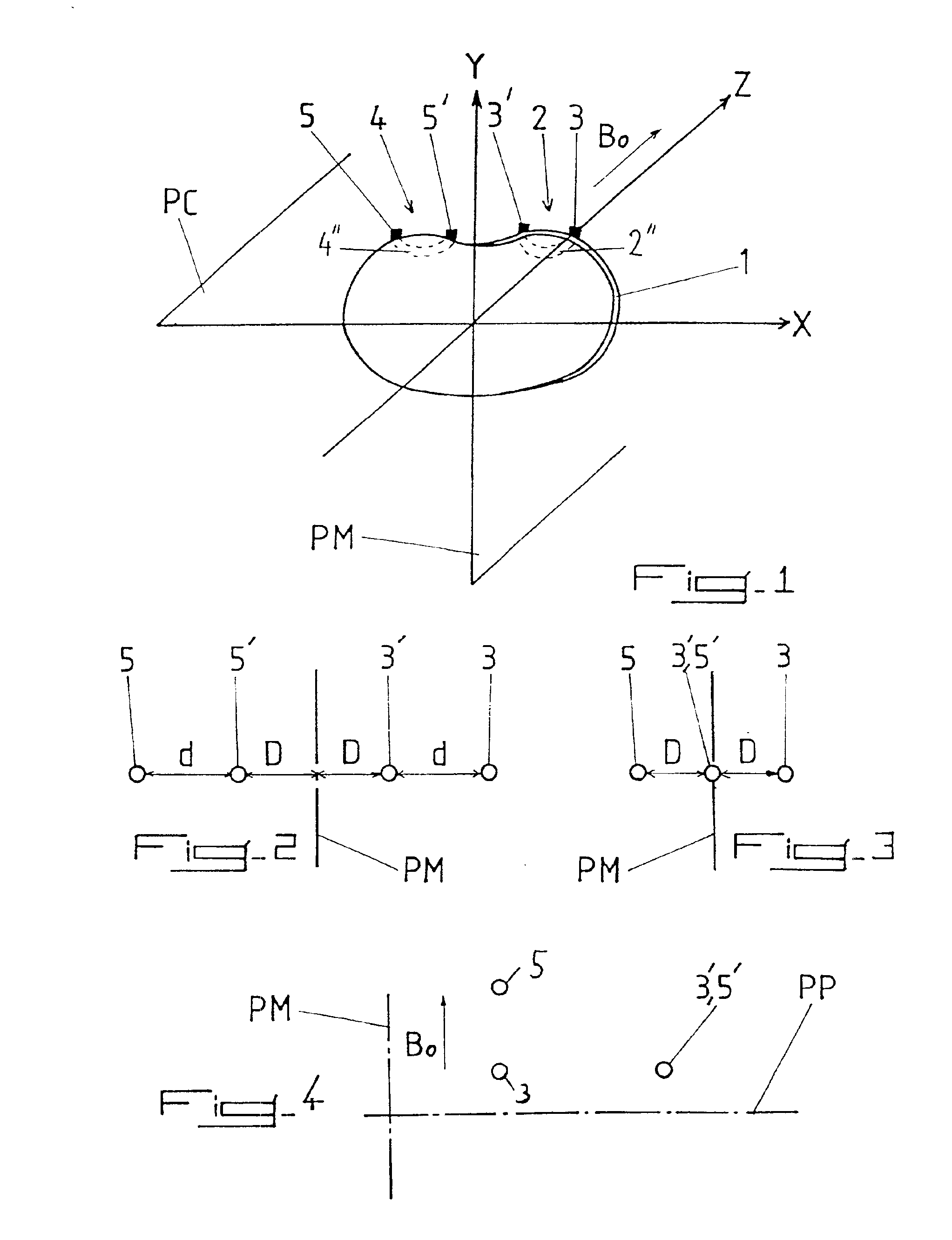
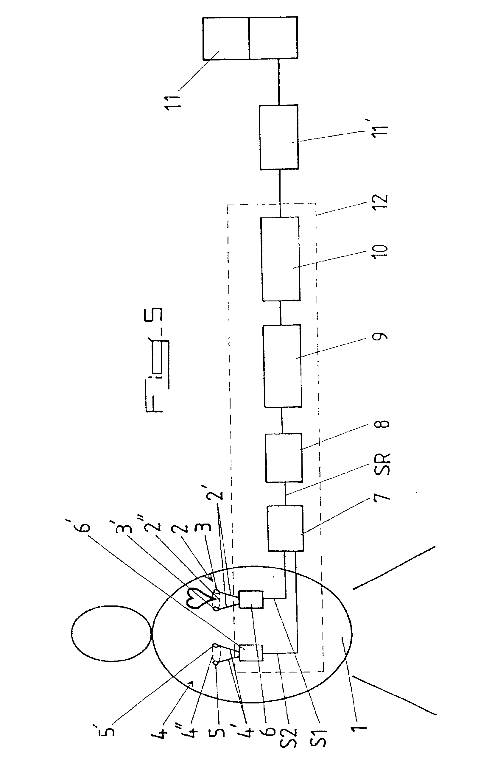
In accordance with the invention, the first and second measurement loops 2 and 4 are disposed essentially symmetrically in relation to the median plane PM of patient 1 perpendicular to the patient's coronal plane PC, while electrodes 3, 3′; 5, 5′ of the two measurement loops 2 and 4 are preferably, but not necessarily, arranged in alignment with similar spacing between the electrodes in each pair 3 and 3′; 5 and 5′ (see FIGS. 1 through 3 and 5 of the attached drawings).

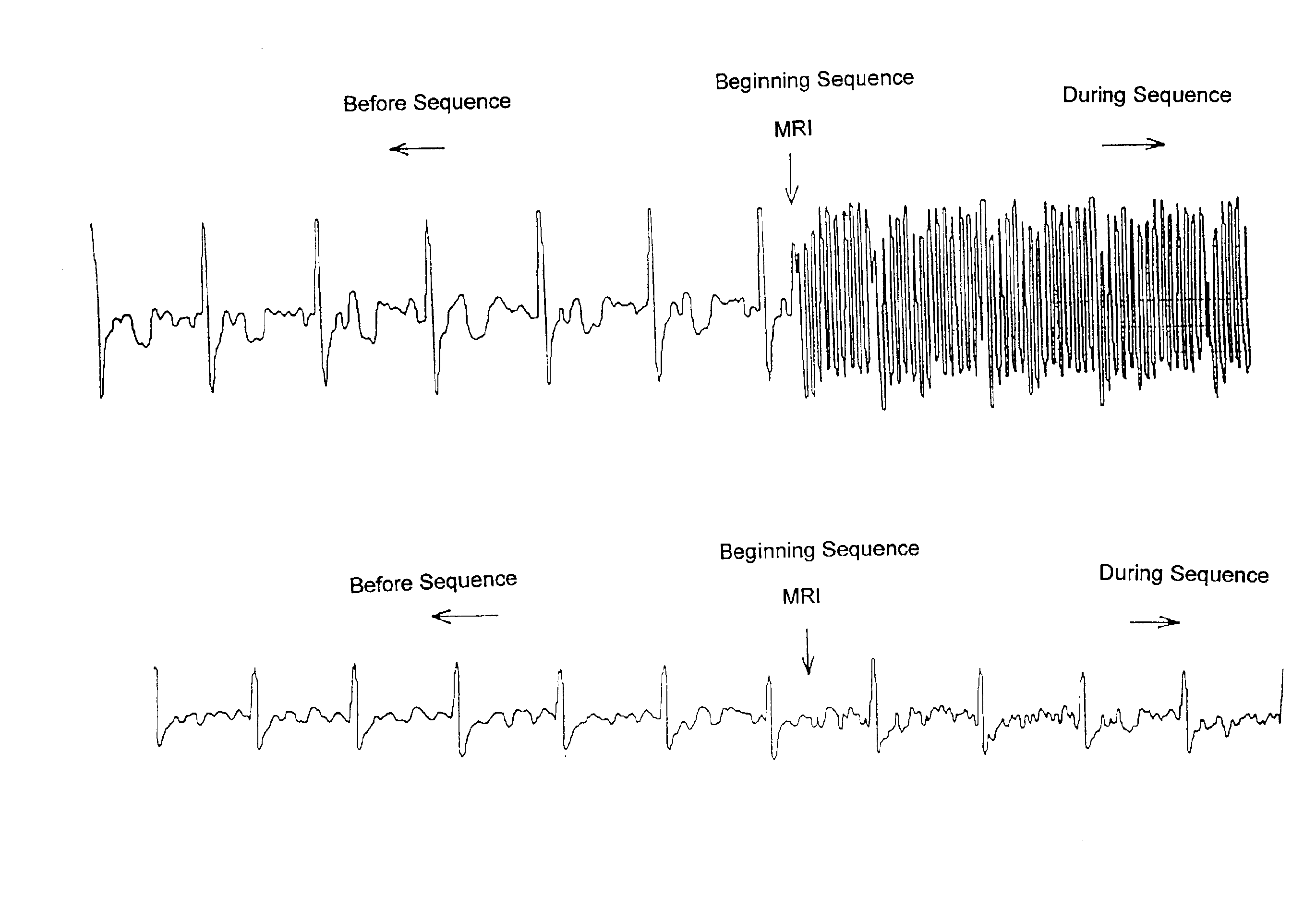
The inventor is able to state with certainty that the sound signals recovered by measurement loops 2 and 4 configured and arranged as described above are essentially identical in current MRI experiments making use of magnetic field gradients.

second embodiment

According to the invention shown in FIG. 4 of the attached drawings, the three electrodes 3, 3′; 5, 5′ forming the two measurement loops 2 and 4 are arranged in a triangular configuration near the cardiac region in such a way that the second measurement loop 4 recovers a signal component representing an ECG with opposite polarity and amplitude essentially equal to the signal component representing the ECG recovered by the first measurement loop 2.

Finally, so that it can be used in real time, especially to monitor the condition of a patient undergoing an MRI examination, the signal SR resulting from the addition or subtraction of the differential signals S1 and S2 delivered by the two measurement loops 2 and 4 can be processed by a pass-through filter 8, then used to modulate the frequency of a carrier and finally, transmitted to an apparatus 11 equipped with a means for displaying the filtered ECG signal SR, possibly after conversion into an optical signal.

It is also possible for th...


PUM


Abstract

A method of obtaining an electrocardiograph (ECG) of a patient located in a turbulent electromagnetic environment. The method includes recovering an ECG signal from a patient. The signal, including added noise, is recovered near the cardiac region as the differential signal resulting from signals delivered by two electrodes forming part of a first measurement loop. The signal further consists of simultaneously recovering from the patient a second measurement signal incorporating at least the noise, as a differential signal resulting from the signals delivered by two electrodes forming part of a second measurement loop distinct from the first measurement loop. Then, the second measurement signal is added or subtracted from the first noisy ECG signal, in real time. The second measurement signal is as a function of the polarity of the noise in the second signal relative to the noise in the first noisy ECG signal. The resulting signal is processed, converted, transmitted and displayed in real time representing the ECG essentially devoid of noise.

Description

The instant invention concerns obtaining and processing physiological signals, especially with a view toward improving the signal / noise relationship, and has as its object a method and a device for capturing an electrocardiograph signal (ECG) which is essentially devoid of noise, particularly noise caused by variations in the local magnetic field.BACKGROUND OF THE INVENTIONIn capturing physiological signals, obtaining a clear signal often poses a problem, as the signal is generally weak and drowned by background noise.This difficulty increases when the signal is acquired in an MRI environment and when the signal involved is an electrocardiograph (ECG).Not only is the electrocardiograph the preferred indicator of a patient's health, but it also serves as a sequencing signal, particularly for an MRI imager; for example, it may function as a signal for launching an acquisition sequence (better known as “triggering”) and / or a signal for launching an acquisition window (better known as “...

Claims


Application Information

Patent Timeline
Patent Type & Authority Patents(United States)
IPC IPC(8): A61B5/0402A61B5/0428A61B5/308
CPCA61B5/0428Y10S128/901A61B5/7285A61B5/30A61B5/28
Inventor FISCHER, ROLAND
Owner SCHILLER MEDICAL
Why Eureka
Try Eureka