Method for fabricating cell-containing implants

a cell-containing and implant technology, applied in the field of cell culture, can solve the problem of not producing sufficient contact between aggregates to produce adequate aggregate siz

Inactive Publication Date: 2005-05-03
CHONDROS +1
View PDF4 Cites 115 Cited by
  • Summary
  • Abstract
  • Description
  • Claims
  • Application Information

AI Technical Summary

Benefits of technology

[0014]Alternatively, the culture-expansion step (Step 3) may be conducted directly in an appropriately designed

Problems solved by technology

Goodwin, because his process is carried out under microgravity conditions, does not produce sufficient contact between aggregates

Method used


Image

  • Method for fabricating cell-containing implants
  • Method for fabricating cell-containing implants
  • Method for fabricating cell-containing implants

Examples


example 1

[0066]This example illustrates that chondrocytes propagated in spinner culture on biopolymer beads eventually aggregate into cartilage-like masses.

[0067]In this experiment, the inventors tested the hypothesis that nasal chondrocytes propagated in microcarrier spinner culture would proliferate and produce extracellular matrix components similar to that produced by articular chondrocytes. Cartilage was obtained from five patients during nasal septum reconstruction. Chondrocytes isolated by collagenase digestion were directly seeded at 4×10³ cells / cm2 onto Cellagen microcarriers (100-400 cm2, derived from bovine corium, (ICN, Cleveland, Ohio)) or in monolayer culture. Monolayer and microcarrier spinner cultures were incubated at 37° C., 5% CO2 for fourteen days. Chondrocytes were harvested and cell samples enumerated in trypan blue vital dye. To analyze for proteoglycans production, cells were pulsed for 60 hours with 50 μCi / ml, 35SO4. The proteoglycans were extracted using 4M guanidiu...

example 2

[0070]Steps (1) and (2) of Example 1 are performed by the same procedures.

[0071]Step (3). The cell-seeded microcarriers are maintained in spinner culture at 60 rpm, 37° C., 5% CO2 for 14 to 21 days to allow visible aggregation to take place in enriched medium (Dulbecco essential media containing NCTC-109, OPI (oxaloacetate, pyruvate, insulin), 1-glutamine, gentamycin, and fetal calf serum). The cell-microcarrier aggregates are subsequently centrifuged at 200 g for 15 minutes and 4° C. The supernatant fluid is removed and the aggregates are resuspended in the same enriched media described above.

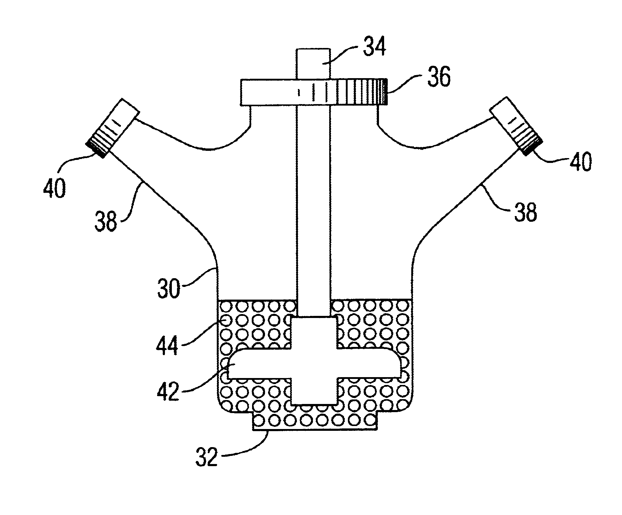
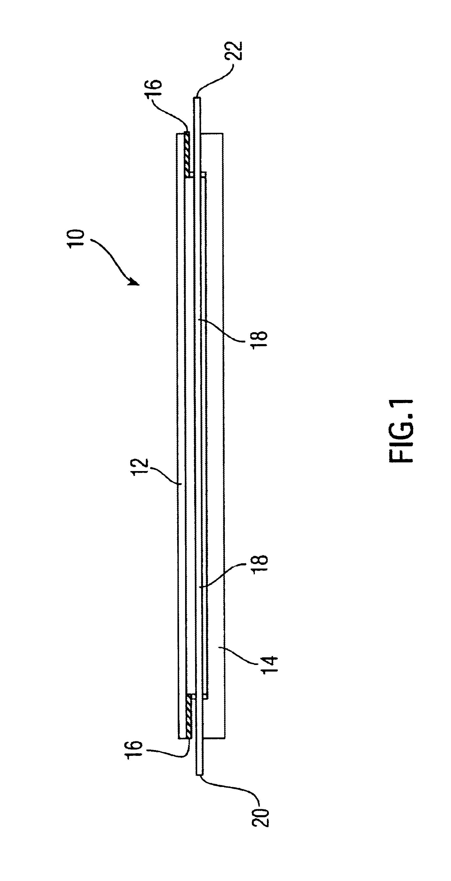
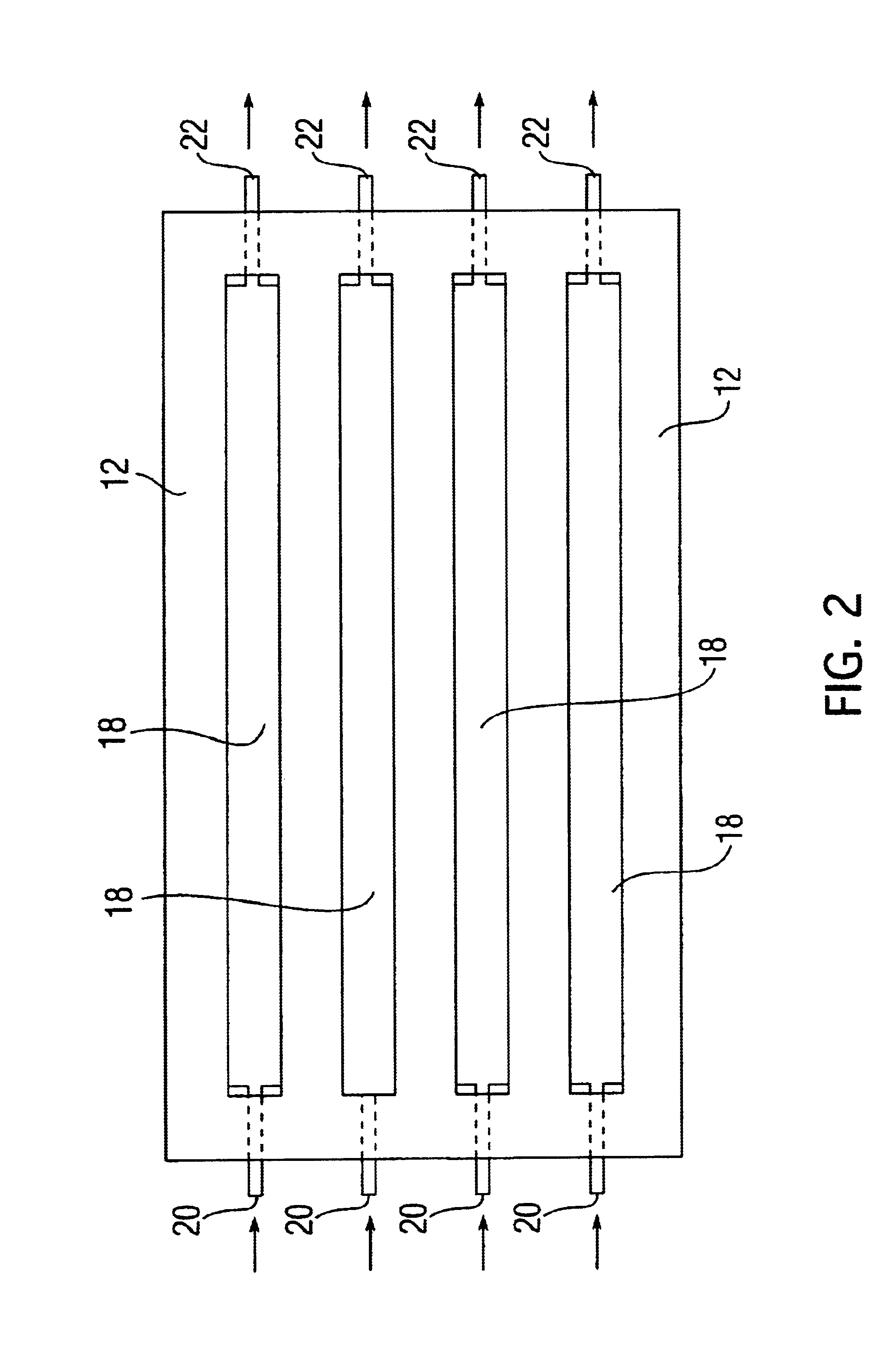
[0072]Step (4). The resuspended aggregates are next transferred by pipetting into the “implant assembly unit (IAU),” as shown in 10. The IAU 10 is siliconized and sterilized before use. The aggregate suspension is transferred into the individualized channel or trough 18 of the “implant assembly unit”. The top lid 12 is then positioned over bottom chamber 14, separated by the rubber gasket 16. ...

example 3

[0076]Steps (1) and (2) of Example 1 are performed by the same procedures.

[0077]Step (3). The cell-seeded microcarriers are maintained in spinner culture at 60 rpm, 37° C, 5% CO2 for 14 to 21 days to allow visible secretion of extracellular matrix to take place in enriched medium (Dulbecco essential media containing NCTC-109, OPI (oxaloacetate, pyruvate, insulin), 1-glutamine, gentamycin, and fetal calf serum). The cell-microcarrier aggregates are subsequently centrifuged at 200 g for 15 minutes and 4° C. The supernatant fluid is removed and the aggregates are resuspended in a fluid medium, such as isotonic saline, phosphate buffered saline solution or Hank's balanced salt solution, suitable for injection into the body.

[0078]Step (4). The resuspended aggregates are next transferred to a syringe or other suitable implantation device whereby the suspension is implanted directly into the anatomic site or cavity requiring the cartilage implant. During the next 14-28 days, or after a sui...


PUM

PropertyMeasurementUnit
Timeaaaaaaaaaa
Timeaaaaaaaaaa
Timeaaaaaaaaaa

Abstract

Disclosed is a method of growing cells on biodegradable microcarrier particles and more specifically growing chondrocytes for an extended period of time until they aggregate. These aggregated cells can be injected directly or shaped for implantation into the body. In another embodiment of this invention, the cell microcarrier aggregates are grown in a mold that is shaped to conform to the geometry of the desired body part to be replaced. An apparatus for shaping the aggregated cells is disclosed. The aggregated cells can be supplied in a kit.

Description

RELATED APPLICATIONS[0001]This application is related to provisional applications Ser. No. 60 / 081,016 filed Apr. 8, 1998 and Ser. No. 60 / 104,842 filed Oct. 20, 1998 and is a continuation-in-part of Ser. No. 09 / 275,319 filed Mar. 24, 1999 now U.S. Pat. No. 6,378,527, and is also related to provisional application Ser. No. 60 / 165,608, filed Nov. 15, 1999, and is also a continuation-in-part of Ser. No. 09 / 712,662, filed Nov. 14, 2000.FIELD OF THE INVENTION[0002]The herein disclosed invention finds applicability in the field of cell culture, as well as in the field of preparing tissue substitutes for tissue replacement and for prosthesis.BACKGROUND[0003]Attempts at replacing or rebuilding diseased or damaged structures in the human body go back to 3000 B.C. It was not until the middle of the 1900's, however, that the use of synthetic materials for rebuilding body structures met with widespread and reproducible success. Advances in material science and biomaterials have afforded much of ...

Claims


Application Information

Patent Timeline
IPC IPC(8): A61K9/16A61K47/36A61K9/50A61L27/38A61L27/00C08B37/02C08B37/00C08B15/00C08B33/00C08J3/12C08H1/00C12N5/00C12N5/06A61K35/12A61F2/08A61K9/00C12N5/077
CPCA61K9/1652A61K9/5036A61K47/36A61L27/3633A61L27/3817A61L27/3895C08B15/005C08B33/00C08B37/00C08B37/0021C08B37/006C08H1/00C08J3/12C12N5/0075C12N5/0654C12N5/0655A61K9/0019A61F2/08C12N2533/72A61K35/12C08J2305/08C12N2500/02C12N2531/00C12N2533/70A61L27/3804
Inventor FRONDOZA, CARMELITA G.FINK, DAVID J.HUNGERFORD, DAVID S.SHIKANI, ALAN H.
Owner CHONDROS
Why Eureka
Try Eureka