Organ-region-indication system incorporated in electronic endoscope system

a technology of electronic endoscopy and region indication, which is applied in the field of electronic endoscopy system, can solve the problems of difficult correct and quick determination of the region of the and it is difficult for even a skilled doctor to correctly and quickly determine the region of the complex organ reproduced on the tv monitor

Inactive Publication Date: 2005-08-16
ASAHI KOGAKU KOGYO KK
View PDF2 Cites 49 Cited by
  • Summary
  • Abstract
  • Description
  • Claims
  • Application Information

AI Technical Summary

Benefits of technology

[0014]Also, preferably, the determining system comprises a numerical-evaluating system that numerically evaluates the degree of coincidence between the read image data and the referential image data, and a comparing system that compares the degree of coincidence with a threshold. When the degree of coincidence is more than the threshold, it is determine

Problems solved by technology

However, the correct and quick determination of the region of the organ reproduced on the TV monitor is very difficult, especially for a novice who manipulates the scope.
Also, when a complex organ, such as the bronchi, is examined using the electron

Method used


Image

  • Organ-region-indication system incorporated in electronic endoscope system
  • Organ-region-indication system incorporated in electronic endoscope system
  • Organ-region-indication system incorporated in electronic endoscope system

Examples


Embodiment Construction

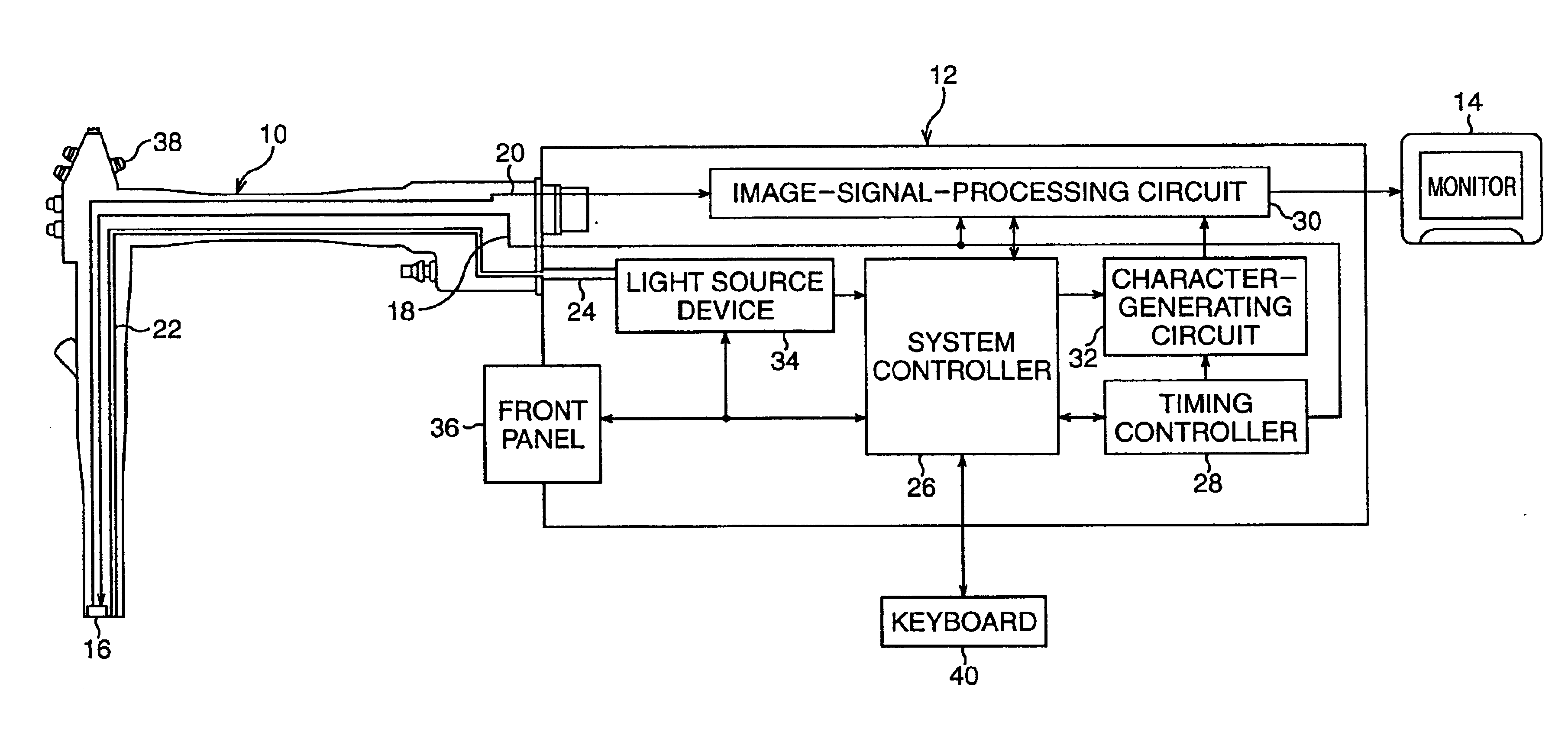
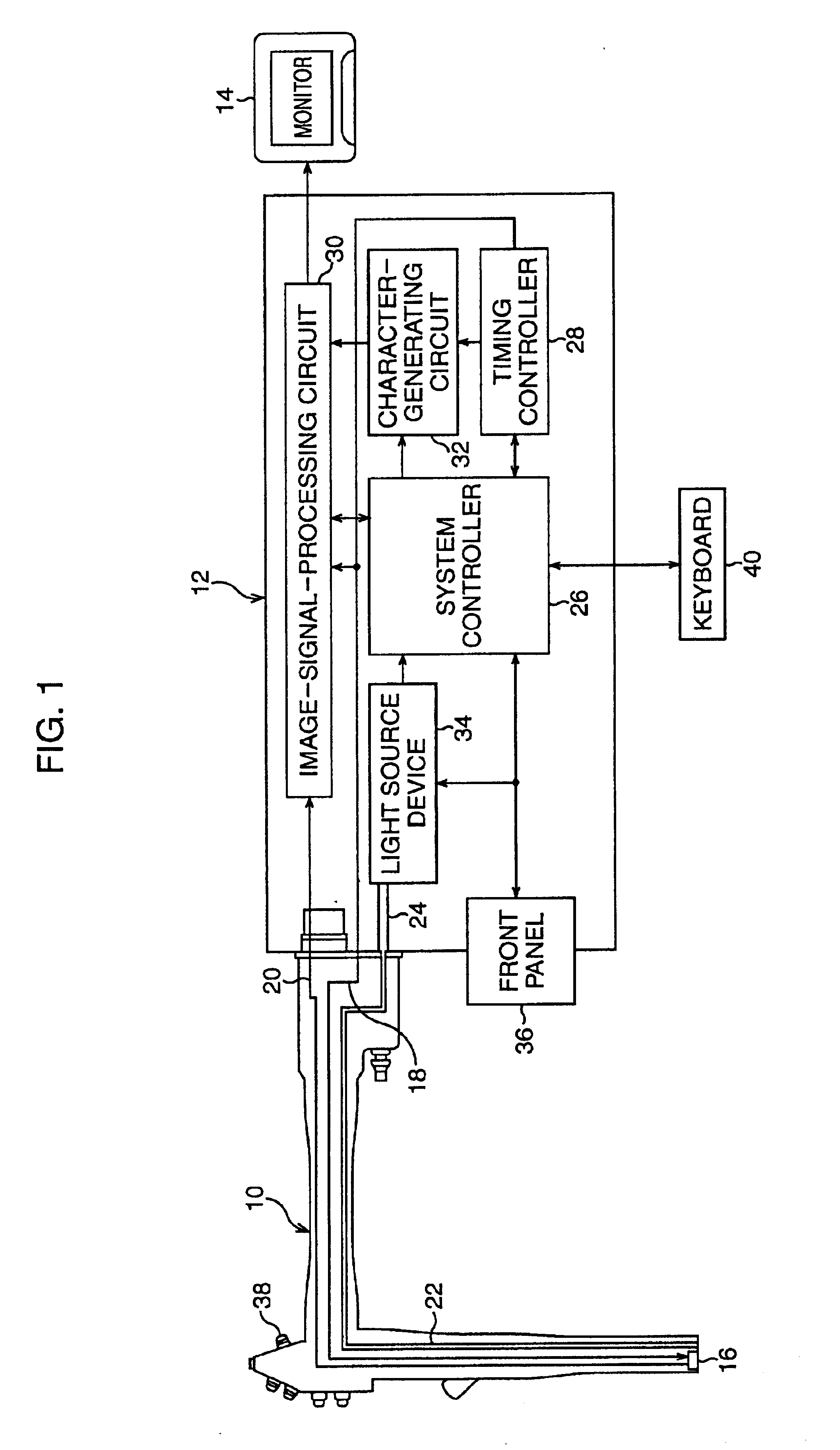
[0033]FIG. 1 schematically shows an electronic endoscope system in which an organ-region-indication system according to the present invention is incorporated. The electronic endoscope system comprises a scope 10, an image-signal processing unit (a so-called processor) 12 to which the scope 10 is detachably coupled, and a TV monitor 14 to which the image-signal-processing unit 12 is connected.

[0034]The scope 10 is representative of various types of scopes, used for bronchial, esophageal, gastro, colon, etc. inspections, and these various types of scopes use the image-signal-processing unit 12 in common. This is because the scope 10 is detachably coupled to the image-signal-processing unit 12.

[0035]The scope 10 is provided with a solid-state image sensor 16, such as a CCD (charge-coupled-device) image sensor, at the distal end thereof, and the CCD image sensor 16 is associated with an objective lens system (not shown). In this embodiment, the CCD image sensor 16 has an on-chip color f...


PUM


Abstract

In an organ-region-indication system incorporated in an electronic endoscope system in which an endoscope image is displayed as a motion image on a monitor in accordance with a video signal produced therein, an organ-region-image data base is formed based on an organ map. A plurality of reference data indicating organ-regions and a plurality of image data representing the organ-region are correspondingly stored in the data base. Still image data is retrieved as referential image data from the video signal at suitable regular time intervals. The data base is searched for image data which coincides with the referential image data after the retrieval of the still image data. Corresponding reference data is displayed on the monitor only when the image data, which coincides with the referential image data, is found by the searching.

Description

BACKGROUND OF THE INVENTION[0001]1. Field of the Invention[0002]The present invention generally relates to an electronic endoscope system, and, in particular, is directed to the development of an electronic endoscope system in which a region of an organ of a human body is indicated during medical examination of the organ using the electronic endoscope system.[0003]2. Description of the Related Art[0004]As is well known, an electronic endoscope system is utilized to medically examine internal organs of a human body, such as, the esophagus, stomach, bronchi, lungs, and so on. In general, the electronic endoscope system includes a scope having a solid-state image sensor provided at a distal end thereof, an image-signal-processing unit to which the scope is detachably coupled, and a TV monitor connected to the image-signal-processing unit.[0005]When the scope is inserted in an internal organ of a human body, the solid-state image sensor optically photographs an endoscope image, and conv...

Claims


Application Information

Patent Timeline
IPC IPC(8): A61B1/04G06F19/00A61B1/00G02B23/24G06T1/00G06T7/00H04N7/18
CPCA61B1/042G06F19/321G16H30/20
Inventor KOBAYASHI, HIROYUKISUGIMOTO, HIDEO
Owner ASAHI KOGAKU KOGYO KK
Why Eureka
Try Eureka