Method and arrangement for medical X-ray imaging

a technology of x-ray imaging and x-ray images, applied in the field of medical x-ray imaging, can solve the problems of insufficient information in the sparse projection data to fully describe the 3-d body, difficult to use a priori information, and insufficient information in the sparse projection data

Inactive Publication Date: 2006-06-27
INSTRUMENTARIUM CORP
View PDF6 Cites 12 Cited by
  • Summary
  • Abstract
  • Description
  • Claims
  • Application Information

AI Technical Summary

Benefits of technology

[0016]The invention is also based on that biological issues have that kind of statistical a priori information that this information can be success fully utilized with Bayesian inversion in medical x-ray imaging. The suitable a priori information makes possible to model the biological tissue mathematically accurately enough and independently of X-ray imaging. From biological tissue it is possible to compile qualitative structural information which makes it possible to utilize the Bayesian method successfully to solve the problems in medical three-dimensional x-ray imaging. There is certain regularity in biological tissues and this regularity is useful especially with the Bayesian method.
[0018]With Bayesian inversion it is possible to utilize a priori information efficiently in 3-D reconstruction from sparse projection data, because the suitable a priori information from the biological tissue makes possible to model the biological tissue mathematically accurately enough and independently of X-ray imaging. In addition, a priori information of the imaging geometry, such as approximate positions of the radiation source and the detector, can be similarly taken into account in a sound, statistical way. Any collection of projection data can be used in the reconstruction. Application-dependent a priori knowledge can be used to regularize the ill-posed reconstruction problem.
[0019]This invention improves the quality of 3-D reconstructions over traditional methods. In addition, the number of radiographs can be minimized without compromising quality of the reconstruction. This is very important in medical applications since the X-ray dose of the patient can be lowered.

Problems solved by technology

Sparse projection data does not contain sufficient information to completely describe the 3-D body.
However, some a priori information about the body is typically available without X-ray imaging.
Traditional reconstruction algorithms such as filtered backprojection (FBP), Fourier reconstruction (FR) or algebraic reconstruction technique (ART) do not give satisfactory reconstructions from sparse projection data.
Reasons for this include requirement for dense full-angle sampling of data, difficulty to use a priori information, for example nonnegativity of the X-ray attenuation coefficient, and poor robustness against measurement noise.
For example the FBP method relies on summing up noise elements with fine sampling, leading to unnecessarily high radiation dose.
Anyway the prior art does not know effective methods to determine the imaging geometry like the motion of the imaging device and / or the object, though all parts of the imaging geometry are rarely precisely known in advance.

Method used


Image

  • Method and arrangement for medical X-ray imaging
  • Method and arrangement for medical X-ray imaging
  • Method and arrangement for medical X-ray imaging

Examples


Embodiment Construction

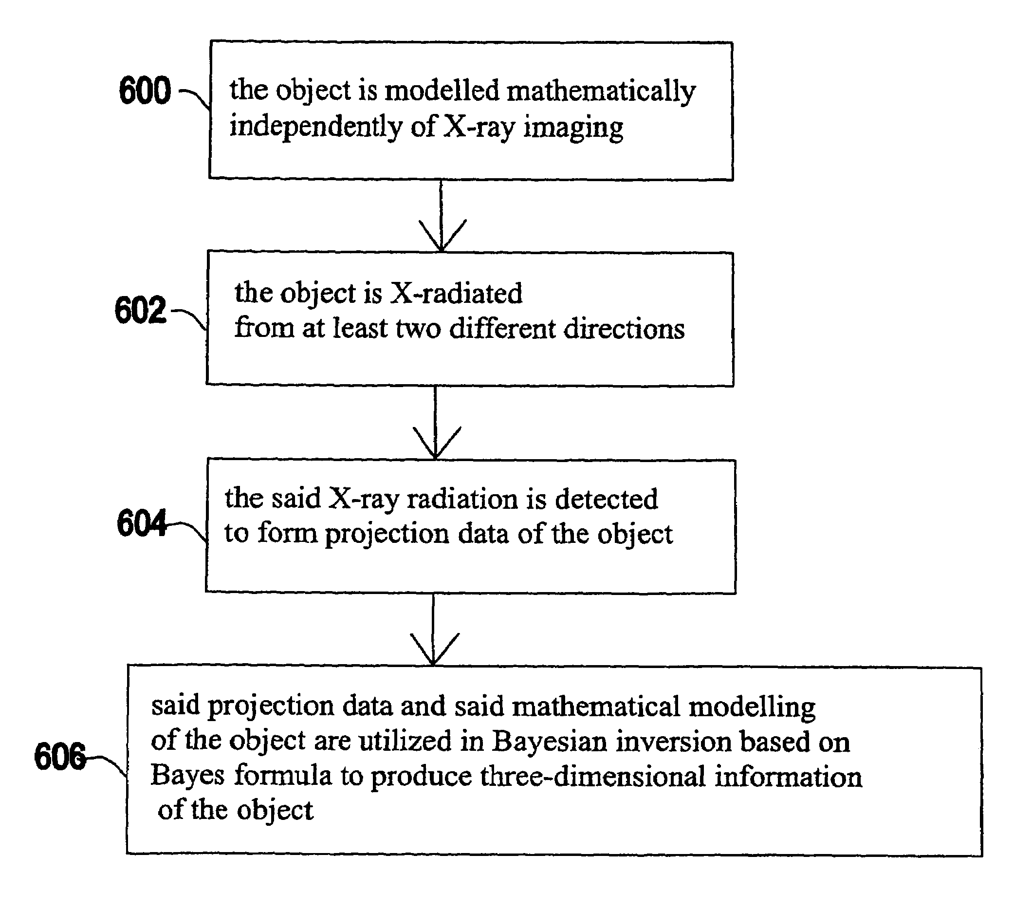
[0029]In conventional tomography, the interior of an object is reconstructed from tomographic projections such as X-ray images. All the current image reconstruction methods assume that projection geometry of the imaging device is either known or solved in advance by using fiducial or non-fiducial feature points in the images. With this invention a novel approach is presented where the imaging geometry is solved with the reconstruction problem while no correspondence information is needed. This approach is a direct application of Bayesian inversion theory and produces the maximum likelihood or maximum a posteriori estimates for the imaging geometry like motion parameters under the selected noise and prior distributions. This invention can be utilized assuming either 1D or 2D projections. To utilize the invention with 1D projections is also often a sufficient assumption since the reconstruction problem is frequently performed as a stack of 2D reconstruction problems for computational ...


PUM

PropertyMeasurementUnit
sizeaaaaaaaaaa
total opening angleaaaaaaaaaa
imagingaaaaaaaaaa

Abstract

A medical X-ray device 5 arrangement for producing three-dimensional information of an object 4 in a medical X-ray imaging comprises an X-ray source 2 for X-radiating the object from at least two different directions; a detector 6 for detecting the X-radiation to form projection data of the object 4; a computational device 15 for modelling the object 4 mathematically utilizing the projection data to solve the imaging geometry and / or the motion of the object, where the solving concerns either some or all parts of the imaging geometry and / or the motion of the object. The computational device 15 utilizes said projection data and said mathematical modelling of the object in Bayesian inversion based on Bayes' formulap⁡(x,θ❘m)=ppr⁡(θ)⁢ppr⁡(x)⁢p⁡(m❘x,θ)p⁡(m)to produce three-dimensional information of the object.

Description

CROSS REFERENCE TO RELATED APPLICATION[0001]The present application claims priority from Finnish Patent Application No. 20031662, filed Nov. 14, 2003.BACKGROUND OF THE INVENTION[0002]Three-dimensional X-ray imaging is based on taking several 1-D or 2-D projection images of a 3-D body from different directions. If 1-D projection images are available from all around a 2-D slice of the body with dense angular sampling, the inner structure of the slice can be determined. This is known as Computerized Tomography (CT) imaging technology, which is widely used in medicine today. A crucial part of CT technology is the reconstruction algorithm taking the X-ray images as argument and producing a voxel representation of the 3-D body.[0003]In many practical cases X-ray projection images are available only from a limited angle of view. A collection of X-ray images of a 3-D body is called sparse projection data if (a) the images are taken from a limited angle of view or (b) there are only a small ...

Claims


Application Information

Patent Timeline
Patent Type & Authority Patents(United States)
IPC IPC(8): A61B6/00G01N23/04G06T11/00
CPCG01N23/046G06T11/006G06T2211/412G01N2223/612Y10S378/901G01N2223/419G06T2211/436
Inventor BRANDT, SAMI
Owner INSTRUMENTARIUM CORP
Why Eureka
Try Eureka