Method and arrangement for medical X-ray imaging and reconstruction from sparse data

a technology of sparse projection data and reconstruction method, applied in image data processing, diagnostics, image data processing details, etc., can solve the problems of insufficient information in the sparse projection data to fully describe the 3-d body, traditional reconstruction algorithms such as filtered backprojection (fbp), fourier reconstruction (fr) or algebraic reconstruction technique (art) do not give satisfactory reconstructions from sparse projection data, and it is difficult to use a priori information

Inactive Publication Date: 2007-09-11
GE HEALTHCARE FINLAND
View PDF2 Cites 38 Cited by
  • Summary
  • Abstract
  • Description
  • Claims
  • Application Information

AI Technical Summary

Benefits of technology

[0014]The invention is based on the fact that biological tissues have that kind of statistical a priori information that this information can be utilized successfully with bayesian inversion in medical x-ray imaging. The suitable a priori information makes it possible to model the biological tissue mathematically accurately enough and independently of X-ray imaging. From biological tissue it is possible to compile qualitative structural information which makes it possible to utilize the bayesian method successfully to solve the problems in medical three-dimensional x-ray imaging. There is a certain regularity in biological tissues and this regularity is useful especially with the bayesian method.
[0016]With Bayesian inversion it is possible to utilize a priori information efficiently in 3-D reconstruction from sparse projection data, because the suitable a priori information from the biological tissue makes possible to model the biological tissue mathematically accurately enough and independently of X-ray imaging. Any collection of projection data can be used in the reconstruction. Application-dependent a priori knowledge can be used to regularize the ill-posed reconstruction problem.
[0017]This invention improves the quality of 3-D reconstructions over traditional methods. In addition, the number of radiographs can be minimized without compromising quality of the reconstruction. This is very important in medical applications since the X-ray dose of the patient can be lowered.

Problems solved by technology

Sparse projection data does not contain sufficient information to completely describe the 3-D body.
However, some a priori information about the body is typically available without X-ray imaging.
Traditional reconstruction algorithms such as filtered backprojection (FBP), Fourier reconstruction (FR) or algebraic reconstruction technique (ART) do not give satisfactory reconstructions from sparse projection data.
Reasons for this include requirement for dense full-angle sampling of data, difficulty to use a priori information for example nonnegativity of the X-ray attenuation coefficient and poor robustness against measurement noise.
For example the FBP method relies on summing up noise elements with fine sampling, leading to unnecessarily high radiation dose.

Method used


Image

  • Method and arrangement for medical X-ray imaging and reconstruction from sparse data
  • Method and arrangement for medical X-ray imaging and reconstruction from sparse data
  • Method and arrangement for medical X-ray imaging and reconstruction from sparse data

Examples


Embodiment Construction

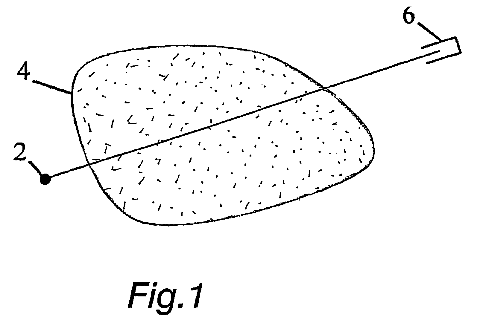
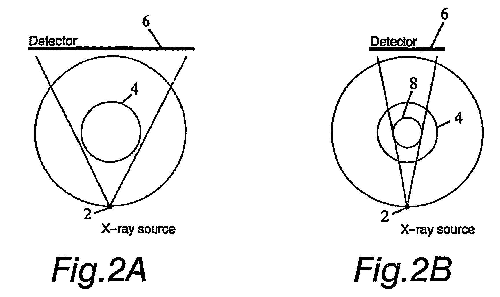
[0027]In practical imaging situations X-ray images are not always available from all around the body. The body might be visible only from certain directions due to imaging geometry. For example this is the case in 3-D mammography with the breast compressed against the detector, or in intraoral dental imaging with the detector inside the patient's mouth. This situation is called limited-angle tomography. Also, the region of interest might be surrounded by tissue that need not be imaged, like in extraoral dental imaging. This situation is called local tomography. In addition the number of radiographs should be minimized in medical applications for reducing the X-ray dose of the patient.

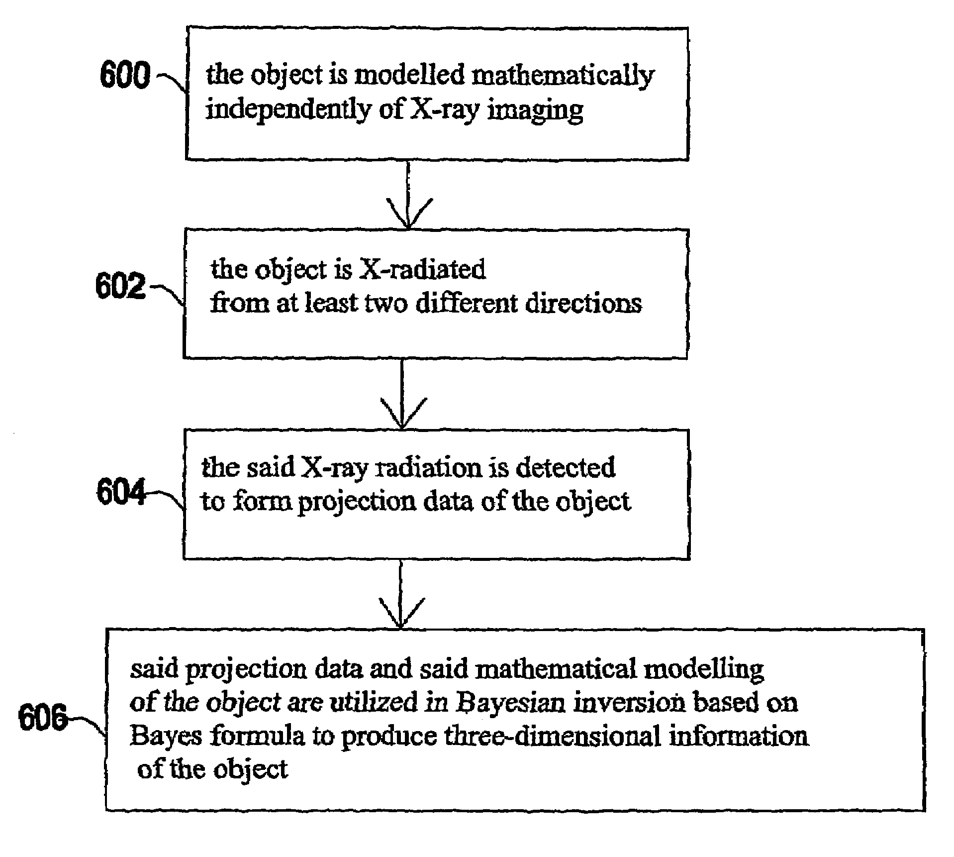
[0028]In the preferred embodiments of the invention Bayesian inversion algorithms are used to create a new type of 3-D medical X-ray imaging. It is intermediate between a projection radiograph and a full computed tomography scan. Two steps are needed to perform successfully such imaging: In step one, th...


PUM

PropertyMeasurementUnit
sizeaaaaaaaaaa
total opening angleaaaaaaaaaa
X-ray aaaaaaaaaa

Abstract

The invention relates to a medical X-ray device 5 arrangement for producing three-dimensional information of an object 4 in a medical X-ray imaging medical X-ray device arrangement comprising an X-ray source 2 for X-radiating the object from different directions and a detector 6 for detecting the X-radiation to form projection data of the object 4. The medical X-ray device 5 arrangement comprises:means 15 for modelling the object 4 mathematically independently of X-ray imagingand means 15 for utilizing said projection data and said mathematical modelling of the object in Bayesian inversion based on Bayes' formulap⁢(⁢x⁢m)=ppr⁡(x)⁢p(m⁢x)p⁡(m)to produce three-dimensional information of the object, the prior distribution ppr(x) representing mathematical modelling of the object, the object image vector x, which comprise values of the X-ray attenuation coefficient inside the object, m representing projection data, the likelihood distribution p(m|x) representing the X-radiation attenuation model between the object image vector x and projection data m, p(m) being a normalization constant and the posteriori distribution p(x|m) representing the three-dimensional information of the object 4.

Description

CROSS REFERENCE TO RELATED APPLICATION[0001]The present application is the U.S. national stage application of International Application PCT / F12003 / 000625, filed Aug. 27, 2003, which international application was published on Mar. 11, 2004 as International Publication WO 2004 / 019782. The International Application claims priority of Finnish Patent Application 20021532, filed Aug. 28, 2002.BACKGROUND OF THE INVENTION[0002]Three-dimensional X-ray Imaging is based on taking several 1-D or 2-D projection images of a 3-D body from different directions. If 1-D projection images are available from all around a 2-D slice of the body with dense angular sampling, the inner structure of the slice can be determined. This is known as Computerized Tomography (CT) imaging technology, which is widely used in medicine today. A crucial part of CT technology is the reconstruction algorithm taking the X-ray images as argument and producing a voxel representation of the 3-D body.[0003]In many practical ca...

Claims


Application Information

Patent Timeline
Patent Type & Authority Patents(United States)
IPC IPC(8): G01N23/00G03B42/02A61B6/00A61B6/02A61B6/03A61B6/14G06T1/00G06T11/00
CPCA61B6/032G06T11/006G06T2211/424G06T2211/436Y10S378/901
Inventor SILTANEN, SAMULISOMERSALO, ERKKIKOLEHMAINEN, VILLELASSAS, MATTIKAIPIO, JARI
Owner GE HEALTHCARE FINLAND
Why Eureka
Try Eureka