
Image-based medical device localization

a medical device and image technology, applied in the field of image-based medical device localization, can solve the problems of difficult to incorporate useful sensors at such small length scales, small diameter, and inability to integrate such specialized sensors or electrodes with every medical devi

Inactive Publication Date: 2007-03-13
STEREOTAXIS
View PDF4 Cites 165 Cited by
  • Summary
  • Abstract
  • Description
  • Claims
  • Application Information

AI Technical Summary

Problems solved by technology

However, incorporating such specialized sensors or electrodes is not always feasible with every medical device.
For instance, thin medical devices such as guide wires are quite small in diameter, and it is difficult to incorporate useful sensors at such small length scales.

Method used


Image

  • Image-based medical device localization
  • Image-based medical device localization
  • Image-based medical device localization

Examples


Embodiment Construction

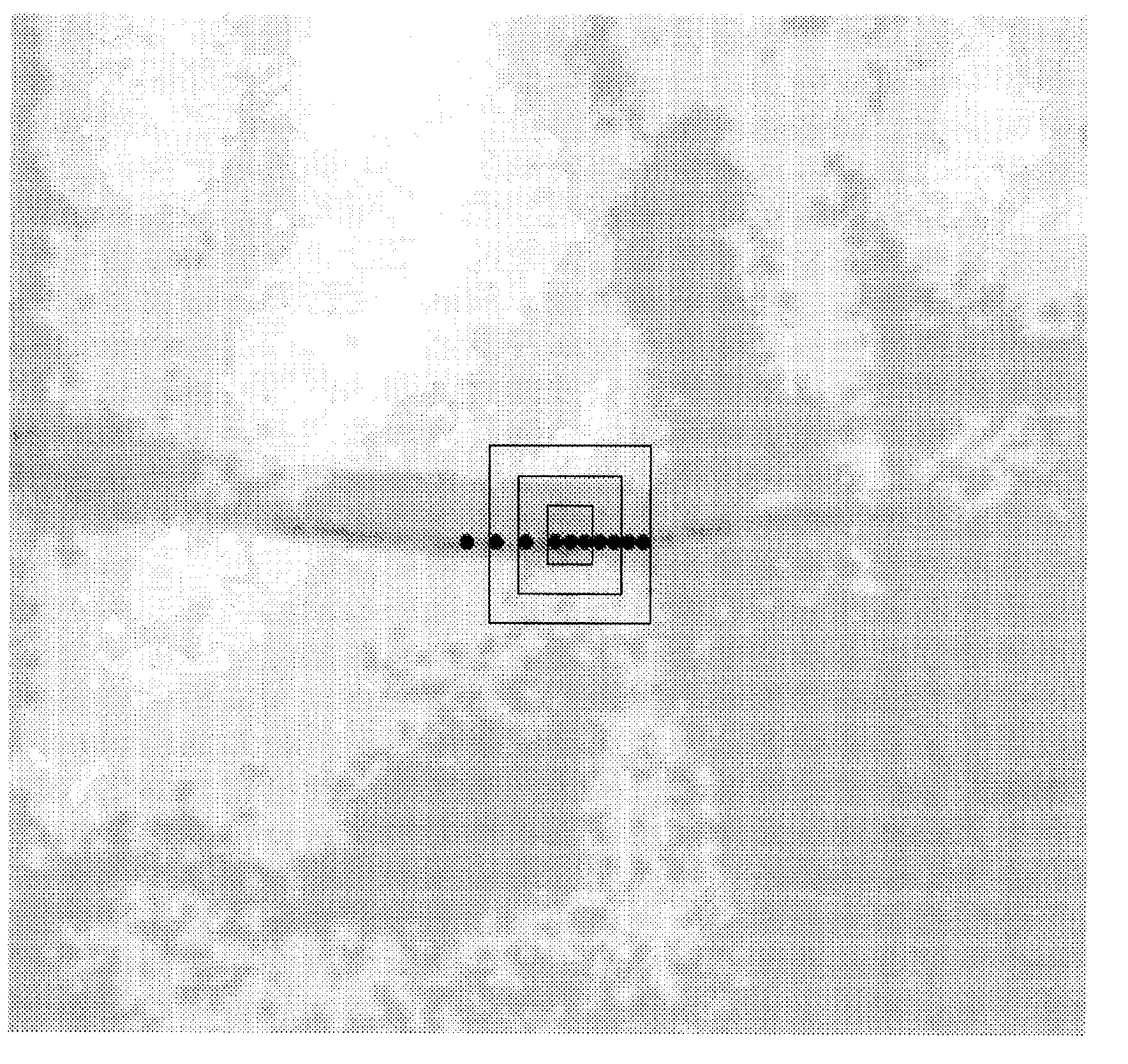
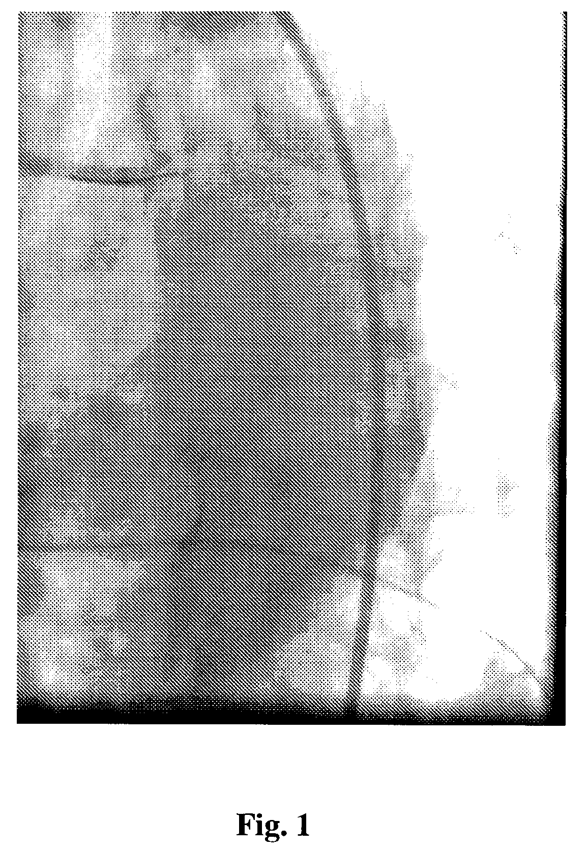
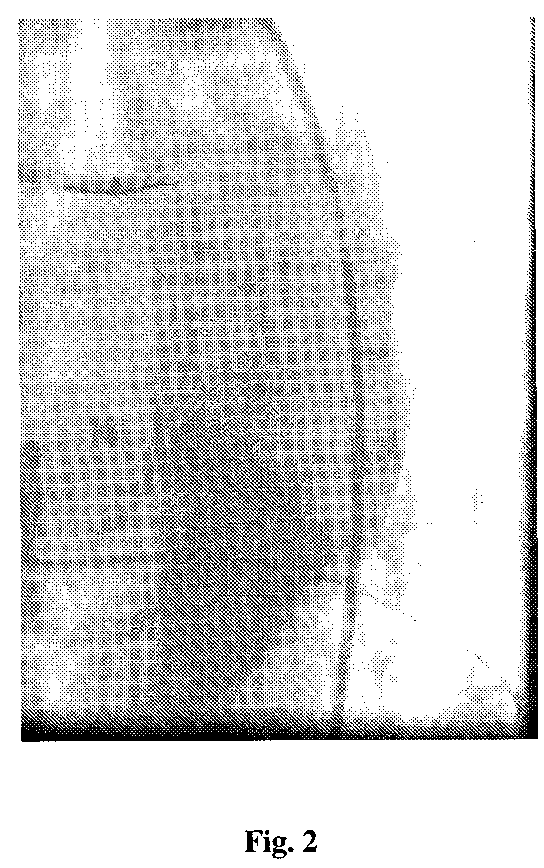
[0012]An elongate medical device is visible on fluoroscopic images when its radio-opacity is sufficiently high, so that it is visible as a darker object against a lighter background. Thus, there is a local contrast difference that is in principle detectable at the level of the pixels that make up the image. For typical elongate devices of the type commonly employed in interventional medical applications, the shape of the device can be thought of as effectively one dimensional. This topological property means that the distribution of darker pixels that make up the fluoro-visible portion of the device can be analyzed to extract the device.

[0013]For example, a medical guide wire can be localized in the three-dimensional space of an operating region in a subject through the image processing of a two dimensional image, such as a fluoroscopic image, of the operating region. While this is invention is described and illustrated in connection with a medical guide wire, the invention is not l...


PUM


Abstract

A method of identifying an elongate medical device in a two-dimensional image of an operating region includes filtering pixels meeting a predetermined intensity requirement for linear scaling, dividing the filtered pixels into groups of contiguous pixels, and selecting from the groups a group corresponding to the medical device.

Description

BACKGROUND OF THE INVENTION[0001]Interventional medical applications have seen a rapid growth in recent years. A variety of techniques and devices for minimally invasive therapies have become commonplace. Of particular note is the emergence of remote navigational systems that offer precise control and steering for medical device navigation. An example of such a system is the Niobe™ Magnetic Navigation System commercialized by Stereotaxis, Inc.[0002]In such a system, the distal tip of the device is remotely actuated from an external User Interface. If the location of the device tip within the anatomy is known, the steering of the device as it is navigated through the anatomy can be automated. Commercially available localization systems such as the CARTO™ EP Navigation System manufactured by Biosense Webster, Inc. can accurately determine the three dimensional location and orientation of a medical device that is equipped with a location sensor. This system requires specialized equipme...

Claims


Application Information

Patent Timeline
Patent Type & Authority Patents(United States)
IPC IPC(8): G06K9/00
CPCG06K9/00G06T7/0012G06T7/403G06T7/0083G06K2209/05G06T2207/30021G06T2207/10121G06T2207/20148G06T7/12G06T7/44G06T7/136G06V2201/03G06F18/00
Inventor VISWANATHAN, RAJU R.
Owner STEREOTAXIS
Learn More