Tomogram creating device and radiation examining apparatus

a tomographic and creating device technology, applied in tomography, applications, instruments, etc., can solve the problems of inability to accurately grasp the swing of each individual region in the body of the examinee, and inability to perform examinations. the effect of reducing the influence of breath-based swing, improving the accuracy of diagnosis of the region on which the radioactive agent is concentrated

Inactive Publication Date: 2007-07-31
HITACHI LTD
View PDF5 Cites 14 Cited by
  • Summary
  • Abstract
  • Description
  • Claims
  • Application Information

AI Technical Summary

Benefits of technology

[0009]An object of the present invention is to provide a tomogram creating device, a tomogram creating method and a radiation examining apparatus capable of improving the accuracy of a diagnosis on a region on which a radioactive medical agent concentrates.
[0011]According to the feature referred to above, the corrections using the respective data about the first structural tomogram obtained based on the first radiation detect signal in the unbreathed state and the second structural tomogram obtained based on the second radiation detect signal in the breathed state make it possible to obtain a tomogram for radiation emitted traceable from a body to be examined, in which the influence of a breath-based swing is less reduced. Therefore, the accuracy of a diagnosis on a region on which a radioactive medical agent is concentrated, is improved.

Problems solved by technology

Since, however, the PET and SPECT or the like are not capable of performing examinations in the unbreathed state, the examinations are performed in a state in which the examinee is being breathed.
Since the examination time becomes long in the method of eliminating the breath artifacts according to the breath synchronization, the same method is inefficient.
Since, however, the correcting method described in Japanese Patent No. 3022773 detects the chest position (breath-based swing) of the examinee by means of the position sensor, the swing of each individual region in the body of the examinee cannot be accurately grasped.
Thus, a tomogram good in accuracy cannot be obtained depending on the correction of Japanese Patent No. 3022773, and the accuracy of a diagnosis effected on a region on which a radioactive medical agent is concentrated in the body, cannot be improved.

Method used


Image

  • Tomogram creating device and radiation examining apparatus
  • Tomogram creating device and radiation examining apparatus
  • Tomogram creating device and radiation examining apparatus

Examples


first embodiment

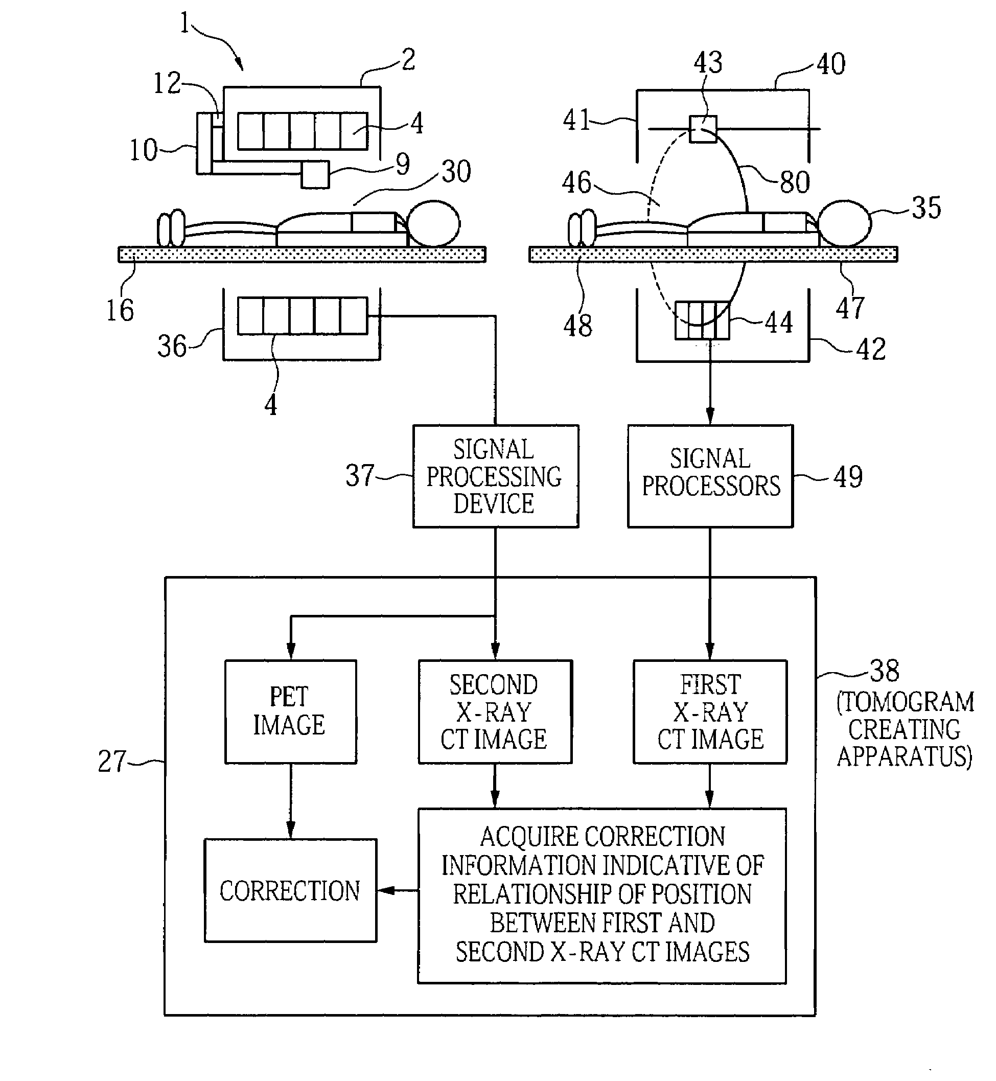
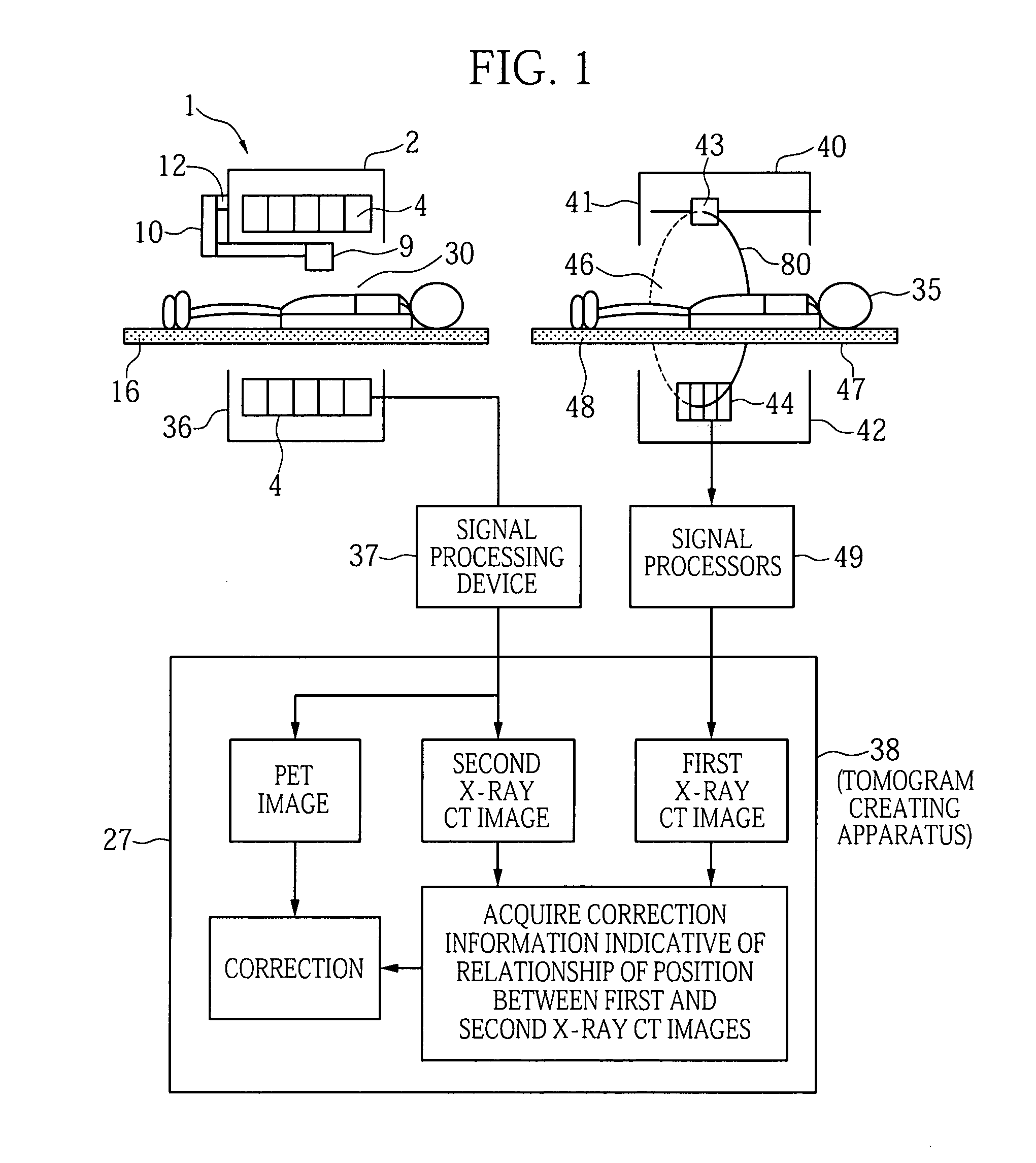
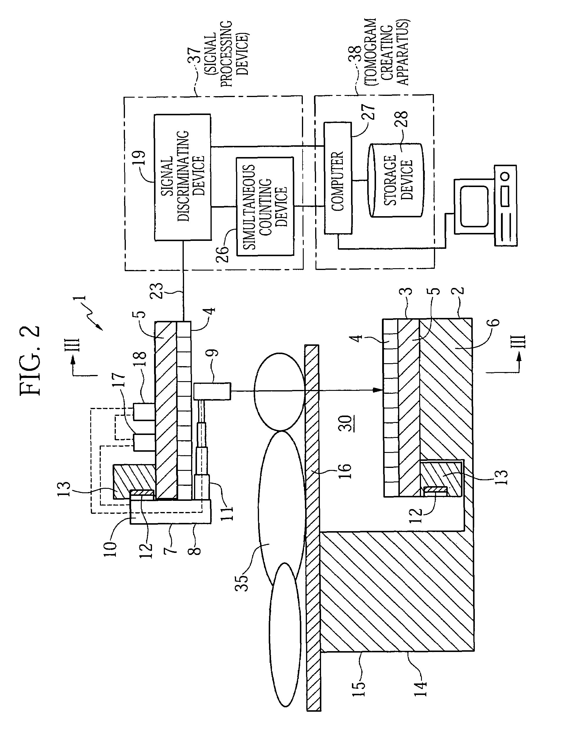
[0024]A first radiation examining apparatus 1 and a second radiation examining apparatus 40 are used in a tomogram creating method showing one preferred embodiment of the present invention as shown in FIG. 1. The radiation examining apparatus 1 is capable of carrying out both a PET examination and an X-ray CT examination (corresponding to a behavior or action for detecting X-rays radiated from an X-ray source and transmitted through a body of an object to be examined or an examinee, by means of a radiation detector) and a PET examination (detecting γ rays radiated from the body due to a radioactive medical agent for PET by means of a radiation detector). The radiation examining apparatus 40 is an X-ray CT for executing an X-ray CT examination.

[0025]A schematic structure of the radiation examining apparatus 40 will be explained below using FIG. 1. The radiation examining apparatus 40 is provided with an imaging device 41 and an examinee holding device 47. The imaging device 41 includ...

second embodiment

[0072]A method of creating tomograms, which shows another embodiment of the present invention, will be explained using FIG. 9. A radiation examining apparatus 1A is used in the tomogram creating method showing the present embodiment. The radiation examining apparatus 1A has a configuration wherein the tomogram creating device 38 of the radiation examining apparatus 1 is replaced by a tomogram creating device 38A. The radiation examining apparatus 1A is identical in other configuration to the radiation examining apparatus 1. The tomogram creating device 38A receives a first X-ray detect signal from its corresponding radiation detector 4.

[0073]In the present embodiment, an X-ray CT examination in an unbreathed state is done using the radiation examining apparatus 1A. Namely, an examinee 35 to which no PET medical agent is administered, is placed on a bed 16 and inserted into a through hole or opening portion 30. An X-ray source 9 is turned around the periphery of the examinee 35 by us...

third embodiment

[0075]A tomogram creating method showing a further embodiment of the present invention will be explained. The radiation examining apparatuses 1 and 40 shown in FIG. 1 are used in the present embodiment. A tomogram creating device 38 employed in the present embodiment executes respective processes of Steps 51 through 53, 55 through 58, 65 through 68 and 61 shown in FIG. 10.

[0076]In a manner similar to the first embodiment, the radiation examining apparatus 40 is used to effect an X-ray CT examination on an examinee 35 in an unbreathed state. The intensity of a first X-ray detect signal outputted from the corresponding radiation detector 44 is determined by a signal processing device 49, which in turn is inputted to a computer 27. An X-ray CT examination and a PET examination in a breathed state are executed using the radiation examining apparatus 1 in a manner similar to the first embodiment. However, the X-ray CT examination in the breathed state is performed in a short period of ti...


PUM


Abstract

An X-ray CT examination using a radiation examining apparatus corresponding to an X-ray CT is effected on an unbreathed examinee. A tomogram creating apparatus creates a first tomogram, based on an X-ray detect signal outputted from a corresponding radiation detector of the radiation examining apparatus. An X-ray CT examination and a PET examination using a radiation examining apparatus are effected on the breathed examinee. The tomogram creating apparatus creates a second tomogram, based on an X-ray detect signal outputted from a corresponding radiation detector of the radiation examining apparatus and creates a third tomogram, based on a γ-ray detect signal outputted from the corresponding radiation detector. Correction information is created based on the first and second tomograms. The third tomogram is corrected using the correction information. Therefore, the corrected third tomogram can be obtained which is not affected by a swing produced with breathing.

Description

BACKGROUND OF THE INVENTION[0001]The present invention relates to a tomogram creating device, a tomogram creating method and a radiation examining apparatus, and particularly to a tomogram creating device, a tomogram creating method and a radiation examining apparatus suitable for application to the creation of tomograms by positron emission CT (Position Emission Computed Tomography (hereinafter called “PET”)) or the creation of tomograms by X-ray CT and single photon emission CT (Single Photon Emission Computed Tomography (hereinafter called “SPECT”)).[0002]As a technology for non-invasively imaging functions, forms in a body of an object to be examined or an examinee, there is known an examination using radiation. As typical ones of radiation examining apparatuses, there are known X-ray CT, MRI, PET, SPECT, etc.[0003]The X-ray CT is a method of irradiating an object to be examined or an examinee with radiation emitted from an X-ray source and imaging the form of the body from tran...

Claims


Application Information

Patent Timeline
Patent Type & Authority Patents(United States)
IPC IPC(8): A61B6/00A61B6/03G01T1/161
CPCA61B6/032A61B6/037A61B6/5235A61B6/03
Inventor KOJIMA, SHINICHIUMEGAKI, KIKUOUENO, YUUICHIROUAMEMIYA, KENSUKEKITAGUCHI, HIROSHIOKAZAKI, TAKASHIYOKOI, KAZUMA
Owner HITACHI LTD
Why Eureka
Try Eureka