
Monoclonal antibody inducing apoptosis

a monoclonal antibody and apoptosis technology, applied in the field of monoclonal antibodies, can solve problems such as elucidating characteristics

Inactive Publication Date: 2009-05-12
CHUGAI PHARMA CO LTD
View PDF32 Cites 22 Cited by
  • Summary
  • Abstract
  • Description
  • Claims
  • Application Information

AI Technical Summary

Benefits of technology

The present invention is about monoclonal antibodies that can recognize and induce apoptosis (cell death) of nucleated blood cells with Integrin Associated Protein (IAP). These antibodies can be used as therapeutic agents for myeloid leukemia and lymphoid leukemia. The invention also provides fragments, peptides, and low molecular weight compounds that can induce apoptosis of nucleated blood cells with IAP. The invention also includes hybridomas that produce the monoclonal antibodies.

Problems solved by technology

However, while one cell line of the splenic stromal cells has been established (CF-1 cells) and its cytological characteristics have been studied, specific antibodies that recognize the surface antigens of these cells have never been prepared, nor have their characteristics been elucidated yet in any way.

Method used


Image

  • Monoclonal antibody inducing apoptosis
  • Monoclonal antibody inducing apoptosis
  • Monoclonal antibody inducing apoptosis

Examples


example 1

Monoclonal Antibody Preparation

(1) Sensitizing Antigen and Immunization Method

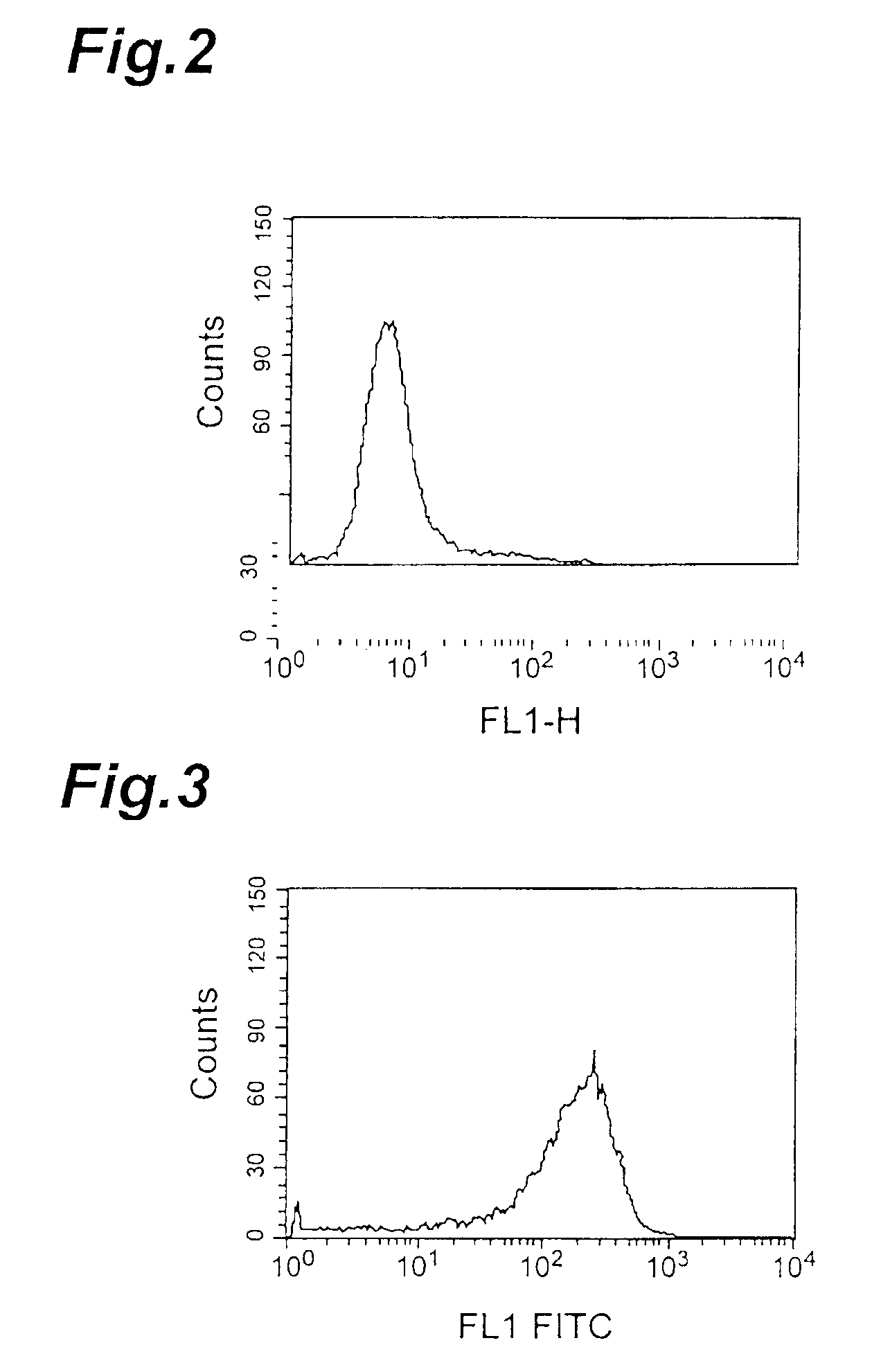
[0079]Antigen sensitization was accomplished using a recombinant cell line as the sensitizing antigen, which was the L1210 cells transfected with human IAP gene and highly expressed the product. L1210 is obtained from the DBA mouse-derived leukemia cell line (ATCC No. CCL-219, J. Natl. Cancer Inst. 10:179-192, 1949).

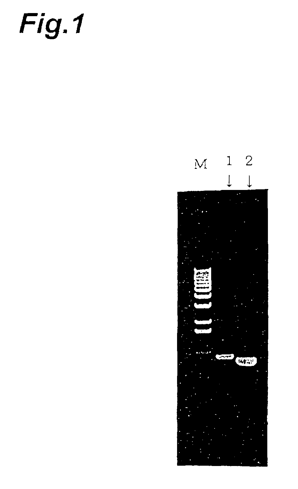
[0080]The human IAP gene was amplified by PCR using a primer with a human IAP-specific sequence (sense primer: GCAAGCTTATGTGGCCCCTGGTAGCG (SEQ ID NO: 1), antisense primer: GCGGCCGCTCAGTTATTCCTAGGAGG) (SEQ ID NO: 2) and cDNA prepared from mRNA of HL-60 cell line (Clontech laboratories, Inc.) as the template (FIG. 1)

[0081]The PCR product was subcloned into a cloning vector pGEM-T (Promega Corporation) and used to transform E. coli JM109 (Takara. Shuzo Co., Ltd.), and after confirming the nucleotide sequence of the insert DNA with a DNA sequencer (373A DNA Sequencer, available from ABI), it was su...

example 2

Subclass Identification of MABL-1 and MABL-2 Antibodies

[0107]In order to identify the subclasses of MABL-1 and MABL-2 antibodies obtained above, 500 μl each of MABL-1 and MABL-2 adjusted to 100 ng / ml was spotted on an Isotyping Kit (Stratagene), by which MABL-1 was shown to be IgG1, κ and MABL-2 was shown to be IgG2a, κ.

example 3

Human IAP-Expressing Human Leukemia Cells

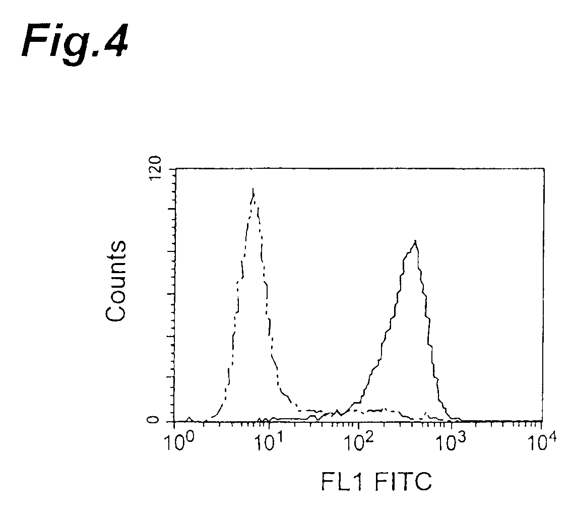
[0108]IAP expression in different human leukemia cell lines was detected by flow cytometry with human IAP-recognizing anti-CD47 antibody (a commercially available product). This antibody was used for the detection because human IAP is believed to be identical to CD47 (Biochem. J., 304, 525-530, 1994). The cell lines used were Jurkat and HL-60 cells (K562 cells, ARH77 cells, Raji cells and CMK cells). The cells were used at 2×10⁵ cells per sample, the anti-CD47 antibody was incubated with the cells at a final concentration of 5 μg / ml, and the secondary antibody used was FITC-labeled anti-mouse IgG antibody (Becton Dickinson and Company). Mouse IgG1 antibody (Zymed Laboratories Inc.) was used as a control. The results of the flow cytometry as shown in FIG. 15 (HL-60) and FIG. 16 (Jurkat) confirmed that both cell lines expressed IAP.


PUM

PropertyMeasurementUnit
concentrationaaaaaaaaaa
temperatureaaaaaaaaaa
concentrationaaaaaaaaaa

Abstract

The monoclonal antibodies of this invention are antibodies that specifically recognize human Integrin Associated Protein, and the antigens that induce apoptosis of nucleated blood cells having human Integrin Associated Protein. Accordingly, they are useful as antibodies that recognize human Integrin Associated Protein for its distinction and identification, while also having an action of inducing apoptosis of nucleated blood cells; these properties can be utilized to prepare useful therapeutic agents in the field of treatment for myeloid leukemia and lymphoid leukemia.

Description

[0001]This application is a continuation-in-part of U.S. patent application Ser. No. 09 / 508,251, filed on Apr. 10, 2000 and now abandoned, which is a national phase application of International Application Serial No. PCT / JP98 / 04118, filed on Sep. 11, 1998,which claims priority to Japanese Patent Application Serial No. 9 / 264853, filed on Sep. 11, 1997. All disclosures are hereby incorporated by reference in their entirety and relied upon.TECHNICAL FIELD[0002]This invention relates to novel monoclonal antibodies having the property of inducing apoptosis of nucleated blood cells with Integrin Associated Protein (IAP) as well as fragments of such monclonal antibodies, peptides or low molecular weight compounds, and to hybridoma that produce the monoclonal antibodies. The novel antibodies are useful as therapeutic agents for myeloid leukemia and lymphoid leukemia.BACKGROUND ART[0003]Granulocyte colony-stimulating factors, such as recombinant granulocyte colony-stimulating factor (rG-CSF)...

Claims


Application Information

Patent Timeline
Patent Type & Authority Patents(United States)
IPC IPC(8): C07K16/00A61K39/395C07K16/28
CPCC07K16/2839A61K2039/505
Inventor FUKUSHIMA, NAOSHIUNO, SHINSUKE
Owner CHUGAI PHARMA CO LTD
Learn More