Image region segmentation system and method

a segmentation system and image technology, applied in the field of medical imaging, can solve the problems of inability to properly segment the desired structure, and the current gray-level segmentation method is not optimal,

Active Publication Date: 2009-09-29
MERATIVE US LP
View PDF8 Cites 36 Cited by
  • Summary
  • Abstract
  • Description
  • Claims
  • Application Information

AI Technical Summary

Problems solved by technology

There are, however, situations in which current gray-level segmentation methods are not optimal.
This characteristic makes it difficult to properly segment the desired structure where distinct structures with similar densities exist.

Method used


Image

  • Image region segmentation system and method
  • Image region segmentation system and method
  • Image region segmentation system and method

Examples


Embodiment Construction

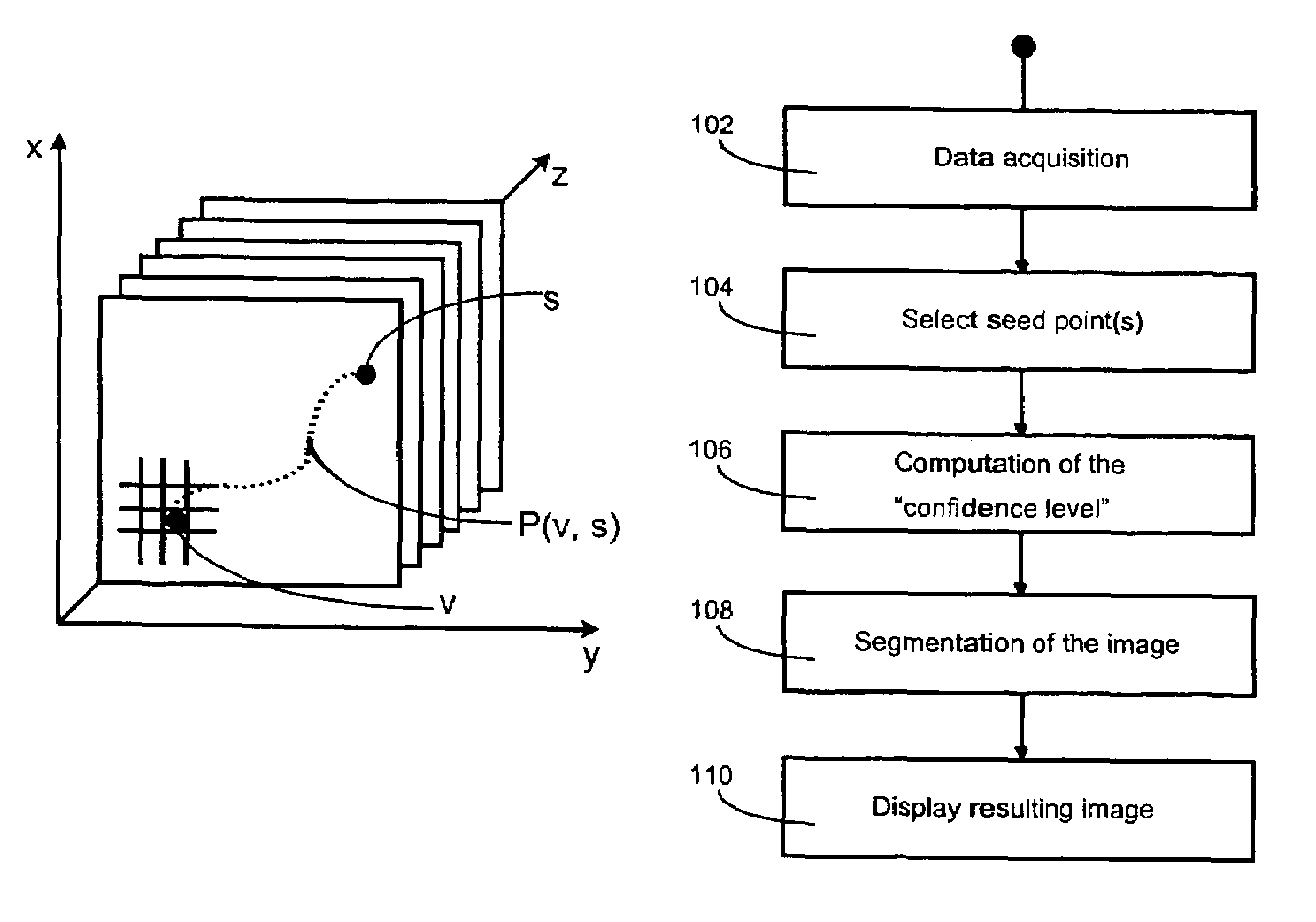
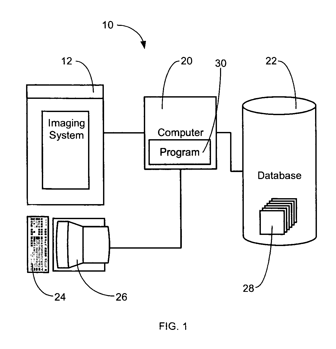
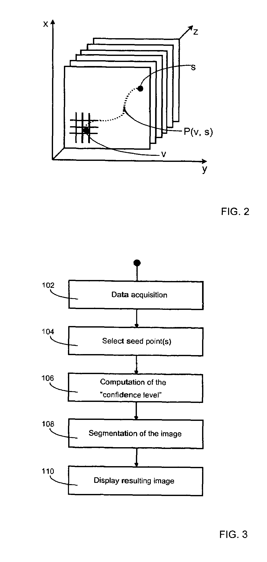
[0026]Referring to FIG. 1, a system for acquiring the image data of a subject, segmenting anatomical structures from the image data and displaying such structures, is indicated generally at numeral 10. Although the embodiment is described with reference to the segmentation of anatomical structures from images obtained by medical imaging systems, it may also be applied to the segmentation of other types of structures from images other than from such systems, such as from, for example, geological survey imaging systems.

[0027]Turning back to FIG. 1, the system 10 comprises a medical imaging system 12 to interrogate a patient and supply data to a computer 20 from which an image can be created. The data is stored as a set of spatially related data points representing variations in a predetermined parameter. Each data point will include information about one or more predetermined parameters than can be displayed to illustrate variations in the parameters. The predetermined parameter is ty...


PUM


Abstract

The invention relates to a method of segmenting an image of a structure stored as a set of spatially related data points representing variations in a predetermined parameter, said method comprising the steps of selecting a seed point within the structure to be segmented, assigning to each of the data points a value of connectivity indicative of the confidence that respective areas of the data points are part of the same structure as said seed point, said value of connectivity including a function of the distance of the respective point from said seed point, establishing a threshold value for said level of connectivity and selecting for display data points meeting said threshold value.

Description

[0001]This application claims priority to U.S. Provisional Patent Application Ser. No. 60 / 450,975 filed on Feb. 28, 2003, the entire contents of which are incorporated herein by reference.[0002]The present invention relates to the field of imaging and in particular to a system and method for segmenting certain subsets of images in order to isolate specific structures.BACKGROUND OF THE INVENTION[0003]In the field of imaging, and more particularly medical imaging, image region segmentation methods are used to isolate subsets of an image corresponding to specific objects such as an artery, the heart, kidneys or any other such anatomical structure. These images may be obtained by various means such as, for example, Magnetic Resonance Imaging (MRI), Computerized Tomography (CT) or any other such medical imaging system. Segmentation, or feature extraction, is an important requirement for any medical imaging application. When a radiologist or a physician looks at an image obtained from a m...

Claims


Application Information

Patent Timeline
Patent Type & Authority Patents(United States)
IPC IPC(8): G06K9/00G06K9/34G06T5/00
CPCG06K9/346G06K9/4638G06T7/0081G06T2207/30101G06T2207/10081G06T2207/20141G06T7/11G06T7/187G06V10/273G06V10/457
Inventor ACCOMAZZI, VITTORIOLAZAROWICH, RENEEBARLOW, CHRISTOPHER J.DAVEY, BRUCE
Owner MERATIVE US LP
Why Eureka
Try Eureka