Method for deriving standard 12-lead electrocardiogram, and monitoring apparatus using the same

a monitoring apparatus and electrocardiogram technology, applied in the field of deriving standard 12-lead electrocardiograms and monitoring apparatuses, can solve the problems of inability difficulty in transmitting a large number of lead waveforms in the form of multi-channel signals, and inability to provide time, etc., to achieve the detection accuracy of the electrocardiogram, achieve the effect of high accuracy, and achieve the effect of easy and effective attainmen

Active Publication Date: 2009-11-03
NIHON KOHDEN CORP
View PDF9 Cites 2 Cited by
  • Summary
  • Abstract
  • Description
  • Claims
  • Application Information

AI Technical Summary

Benefits of technology

[0019]It is therefore an object of the invention to provide a method and a monitoring apparatus for deriving a standard 12-lead electrocardiogram which enables derivation of a standard 12-lead electrocardiogram easily, conveniently and accurately through the use of a minimum number of electrodes; and which is capable of easily and effectively attaining monitoring for appropriate diagnosis and treatment of a variety of heart diseases.
[0029]With the above configuration, the positions of the first electrodes can be identified very easily. In addition, since the respective electrodes are located on an electrical lead line connecting two wrists and the heart, it is possible to reduce an error between the results obtained by the invention and the original limb leads of the standard 12-lead electrocardiogram.
[0030]In addition, since the position of the lead V2 is located at the fourth intercostal left sternal border, and that of the lead V4 is located at an intersecting point of the fifth intercostal and the mid-clavicle line, the positions for the second electrodes are positions that can be most easily identified among the chest leads. Furthermore, a space for attaching a pad for applying defibrillation on the patient suffering ventricular fibrillation can be ensured. Moreover, since an overall reduction is achieved in a sum of electrical angles formed with a direction of the heart vector and the lead vectors of unknown leads V1, V3, V5, and V6, in addition to the leads I and II measured from the limb leads, an error can be minimized.
[0033]In a case where lead vectors of unknown leads V1, V3, V5, and V6 for use in obtaining a heart vector are determined on the basis of a position of the heart and electrode positions of a model of an average build, magnitude of those lead vectors; that is, sensitivity at a time when an instantaneous electromotive force of the heart is transmitted through a living body and appears on a surface of the body can be defined in an optimized manner on the basis of electrical angles formed with lead vectors of unknown leads and lead vectors of leads I, II, V2, and V4 for use in calculation of the third electrocardiogram data set. Accordingly, an error in synthesis of an electrocardiogram due to the build of the patient, attachment positions of electrodes, or positional offset of the heart resulting from body position change can be minimized.
[0043]With this configuration, highly-accurate electrocardiogram data corresponding to the standard 12-lead electrocardiogram set can be obtained easily with attachment of a minimum number of electrodes without requiring rich experience, thereby easily and effectively attaining monitoring for conducting appropriate diagnosis and treatment of a variety of heart diseases.

Problems solved by technology

However, when at-home or emergency medical treatment is to be performed, no time is available for attaching a large number of electrodes to appropriate positions on the body surface of the living body, from the viewpoint of the status of the patient; moreover, difficulty is encountered in transmitting a large number of lead waveforms in the form of multi-channel signals.
However, when leads from the four special positions on the chest surface of the living body are emplaced, appropriate attachment of the electrodes to respective specified positions encounters difficulty, since health care professionals, such as doctors and nurses, are not clinically accustomed to this attachment work, thereby posing a problem of variation arising in detection accuracy of the electrocardiogram.
Furthermore, since none of the leads are actually measured values of the 12 leads, some doubts arise with regard to reliability.
Therefore, accuracy in positioning to the specified positions in attachment of the electrodes poses considerable influences to a measurement result which is inconvenient, in that the attachment requires rich experience, and the like.
Accordingly, problems arise not only in terms of inconvenience for the patient, but also in terms of increased load on a health care professional who applies the electrodes.
This offset can result in an error of lead vectors for calculation of an electrocardiogram derived from unknown leads.
In such a situation, since the lead potential closes to zero, probability of an error in calculation of the heart vector may be increased.

Method used


Image

  • Method for deriving standard 12-lead electrocardiogram, and monitoring apparatus using the same
  • Method for deriving standard 12-lead electrocardiogram, and monitoring apparatus using the same
  • Method for deriving standard 12-lead electrocardiogram, and monitoring apparatus using the same

Examples


Embodiment Construction

[0051]Embodiments of the present invention will be described below in detail with reference to the accompanying drawings.

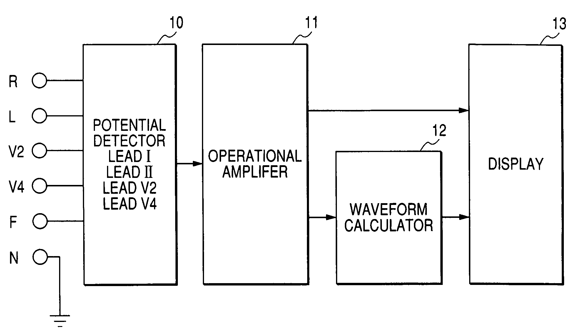
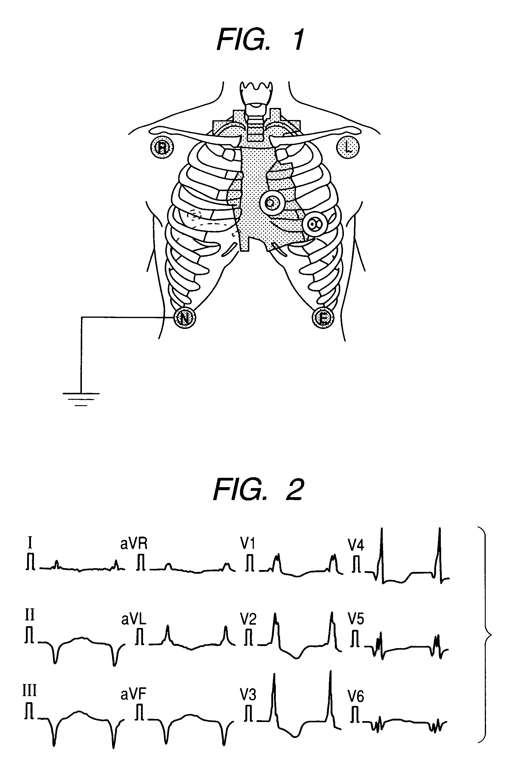
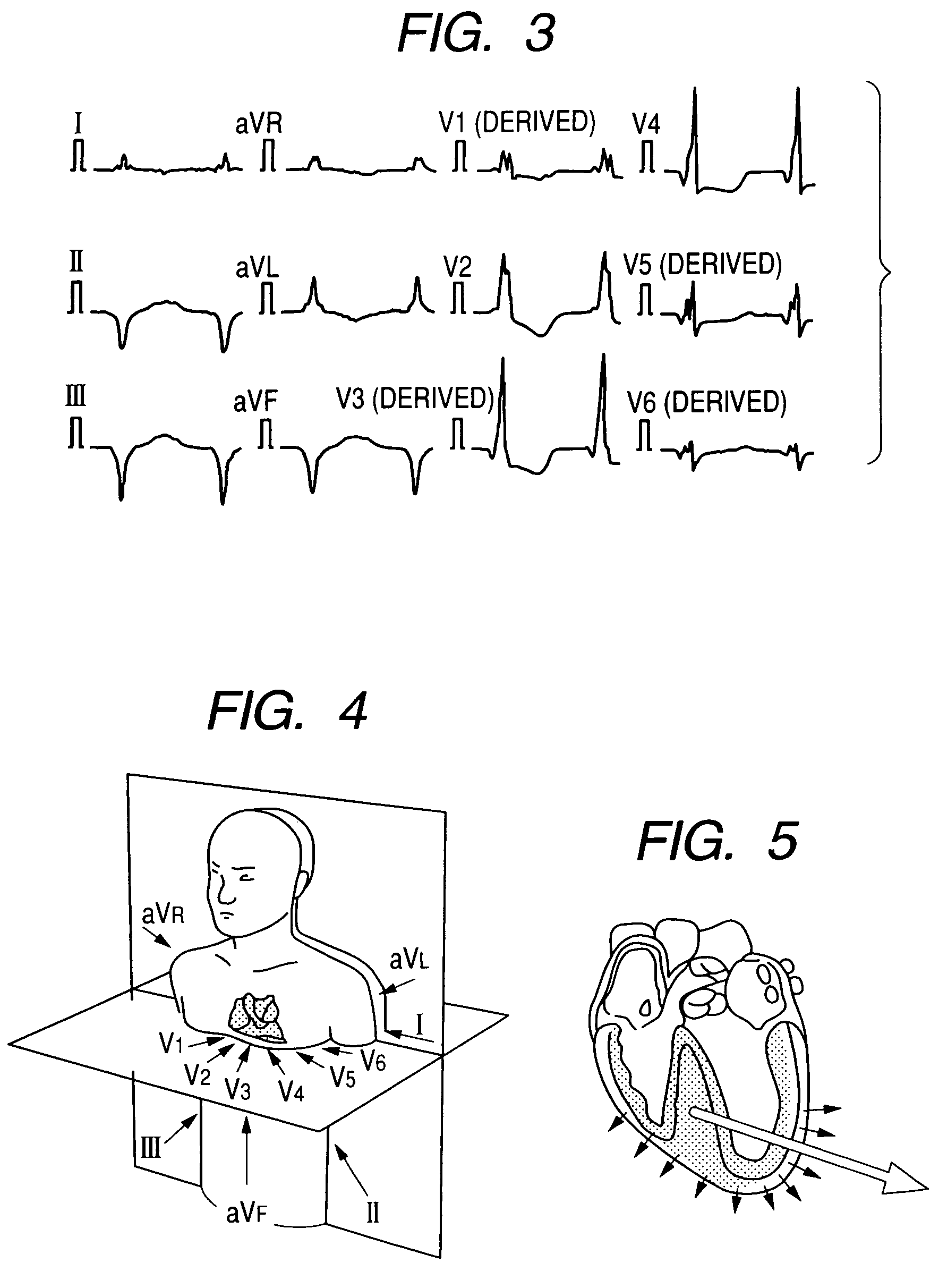
[0052]A rationale for deriving a 12-lead electrocardiogram according to the invention is as follows. In a clinical electrocardiogram a standard 12-lead electrocardiogram is derived by detecting potentials on the basis of Table 1 in accordance with a lead theory. According to the theory, as shown in FIG. 5, an instantaneous electromotive force of the heart at an arbitrary time can be expressed by dipoles at fixed positions; and a potential (V) at an arbitrary lead position can be calculated from the following equation.

V=L·H  (1)

where V denotes a potential matrix, H denotes a heart vector, and L denotes a lead vector.

[0053]The lead vector L of a person was measured by Frank as early as 1953 with use of an artificial human-body model, and this measurement has been recognized to be effective as a foundation of the current vector electrocardiogram. In addition, since t...


PUM


Abstract

Four first electrodes are attached on right clavicle, the vicinity of on left clavicle, on right lowermost rib, and the position on left lowermost rib, corresponding to limb leads 12-lead electrocardiogram (ECG). Two second electrodes are attached on such positions of the living body that correspond to a lead V2 and a lead V4 of 12-lead ECG. First ECG data set correspond to leads I and II of 12-lead ECG. A second ECG data set including the leads V2 and V4. A heart vector is calculated on the first and second ECG data sets, and predetermined first lead vectors of leads I, II, V2 and V4. A third ECG data set including leads V1, V3, V5 and V6 is calculated based on the heart vector and predetermined second lead vectors of leads V1, V3, V5 and V6. A fourth ECG data set from leads III, aVR, aVL and aVF of 12-lead ECG based on the first ECG data set. The 12-lead ECG is derived based on the first to fourth ECG data sets.

Description

BACKGROUND OF THE INVENTION[0001]The present invention relates to a method for deriving a standard 12-lead electrocardiogram effective for diagnosing ischemic heart disease, acute myocardial infarction, or the like, in which a minimum number of electrodes are attached to predetermined areas on the body surface of a living body. The present invention also relates to a monitoring apparatus using such a method.[0002]Conventionally, when an electrocardiogram of a patient is detected, measured, and recorded in a hospital or a like facility, a total of ten electrodes are attached to the body surface of the patient; namely, six positions for chest leads, and four positions for limb leads. Six limb-lead waveforms (I, II, III, aVR, aVL, and aVF) of standard 12-lead waveforms and six chest-lead waveforms (V1, V2, V3, V4, V5, and V6) of the same are derived from electric potentials of the heart detected and measured by the ten electrodes by measuring means, such as an electrocardiograph.[0003]...

Claims


Application Information

Patent Timeline
Patent Type & Authority Patents(United States)
IPC IPC(8): A61B5/04A61B5/308
CPCA61B5/0402A61B5/04028A61B5/0428A61B5/327A61B5/30A61B5/318
Inventor WEI, DAMINGSAKAI, YOSHIO
Owner NIHON KOHDEN CORP
Why Eureka
Try Eureka