Autologous wound sealing apparatus

a technology of autologous wounds and sealing devices, applied in the field of autologous wound sealing apparatus, can solve the problems of presenting numerous problems, reducing the safety of patients,

Inactive Publication Date: 2010-08-10
ABBOTT LAB VASCULAR ENTERPRISE
View PDF12 Cites 16 Cited by
  • Summary
  • Abstract
  • Description
  • Claims
  • Application Information

AI Technical Summary

Benefits of technology

[0013]It also is an object of the present invention to provide apparatus for sealing a puncture tract that are easy to use, and decrease opportunities for error and contamination.
[0014]It further is an object of the present invention to provide apparatus for sealing a puncture tract that facilitate placement of the apparatus relative to a vessel.
[0015]It even further is an object of the present invention to provide apparatus for sealing a puncture tract that prevent leakage of blood congealing agents into a vessel during delivery thereof.
[0020]Disposition of the autologbus plug formed from the coagulated blood into the puncture tract seals the puncture tract and vessel from leakage. The tissue surrounding the puncture tract compressively engages the autologous plug along its length, generating frictional forces that prevent the plug from becoming dislodged into the vessel.

Problems solved by technology

This method, however, presents numerous problems.
In addition, there exists a risk of hematoma since bleeding from the puncture may continue until sufficient clotting occurs, particularly if the patient moves during the clotting process.
Furthermore, application of external pressure to stop bleeding may be unsuitable for patients with substantial amounts of subcutaneous adipose tissue since the skin surface may be a considerable distance from the puncture site, thereby rendering external compression less effective.
This method, however, often requires a complex procedure and requires considerable skill by the medical practitioner.
A significant drawback to the Fagan device is that, when deployed into a vessel, the device may protrude into the blood stream, thereby disturbing blood flow and causing thrombosis in the vessel.
Such apparatus may be unsuitable for some patients due to an adverse immunological reaction to animal-derived collagen, which could lead to anaphylactic shock.
Disadvantageously, the apparatus described in that patent comprises a multiplicity of primarily standard, off-the-shelf components that a medical practitioner would have to assemble prior to use.
This greatly is complicates the procedure, and increases opportunities for human error and contamination.
Furthermore, the apparatus resulting from the assembly of the numerous individual components may be unwieldy to use and expensive.

Method used


Image

  • Autologous wound sealing apparatus
  • Autologous wound sealing apparatus
  • Autologous wound sealing apparatus

Examples


Embodiment Construction

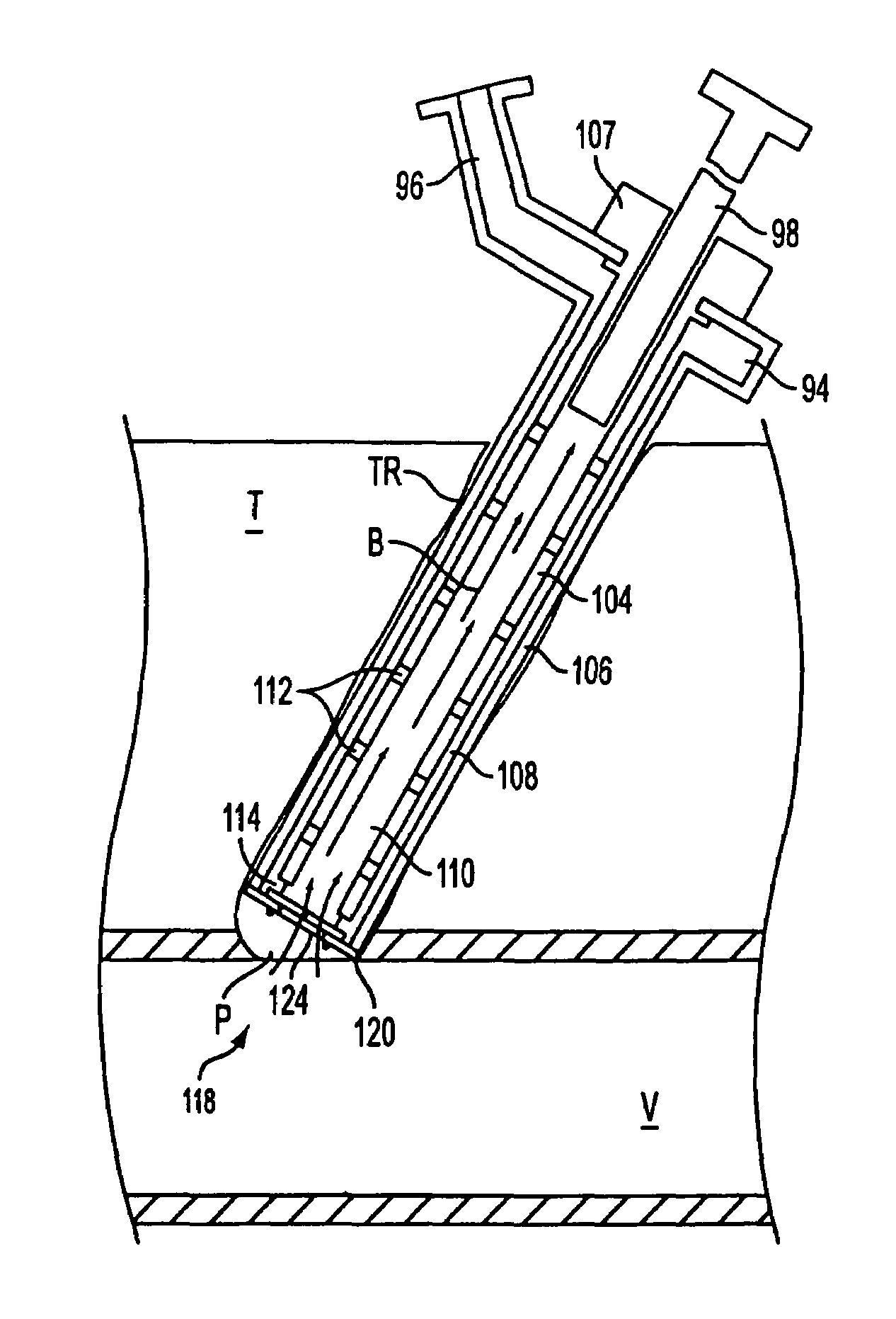
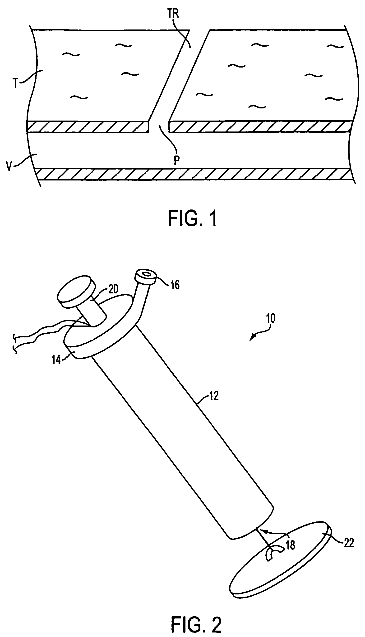
[0035]Upon completion of a medical diagnostic or therapeutic procedure involving percutaneous introduction of instrumentation into blood vessel V, removal of the instrumentation from the patient leaves puncture tract TR. As seen in FIG. 1, puncture tract TR extends through subcutaneous tissue T and terminates at puncture P. The apparatus of the present invention is directed to a device for sealing puncture tract TR by facilitating formation and disposition of an autologous plug within the puncture tract. More specifically, the apparatus facilitates formation of the plug by drawing blood into a lumen of the apparatus, and providing a blood congealing agent to the blood therein, which causes the blood to clot and form an autologous plug within the lumen. The autologous plug is extruded from the lumen to seal puncture tract TR, thereby sealing vessel V from blood leakage.

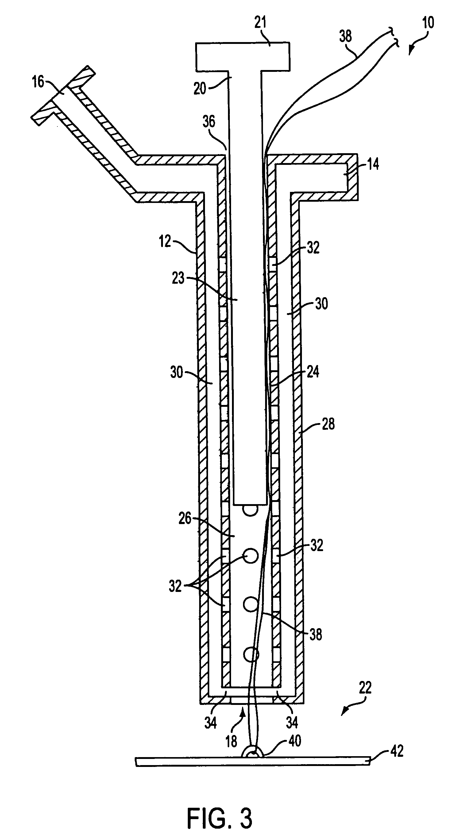
[0036]An illustrative embodiment of device 10 of the present invention is shown in FIGS. 2 and 3. Device 10 comprise...


PUM


Abstract

Apparatus (10) is provided for sealing a vascular puncture tract by forming the autologous plug within the puncture tract, and extruding that plug into the puncture tract. The apparatus of the present invention forms an autologous blood plug by drawing blood into the apparatus from a vessel, mixing a blood congealing agent with the drawn blood, and ejecting a plug formed from the clotted blood within the puncture tract. Also provided are various closure elements (22) to isolate the drawn blood from the vessel during mixture with the blood congealing agent, and to facilitate placement of the apparatus relative to the vessel.

Description

FIELD OF THE INVENTION[0001]The present invention relates to apparatus for sealing puncture tracts. More specifically, the invention relates to apparatus that seals a puncture tract by forming and extruding an autologous plug therein.BACKGROUND OF THE INVENTION[0002]A large number of medical diagnostic and therapeutic procedures involve the percutaneous introduction of instrumentation into the blood vessel. For example, coronary angioplasty, angiography, atherectomy, stenting, and numerous other procedures often involve accessing the vasculature through placement of a catheter or other device in a patient's femoral artery or other blood vessel. Once the procedure is completed and the catheter or other diagnostic or therapeutic device is removed, bleeding from the resultant vascular puncture must be stopped.[0003]Traditionally, a medical practitioner applies external pressure to the puncture site to stem bleeding until hemostasis occurs (i.e. when the clotting and tissue rebuilding h...

Claims


Application Information

Patent Timeline
Patent Type & Authority Patents(United States)
IPC IPC(8): A61B17/08A61B17/00A61B17/12
CPCA61B17/00491A61B17/0057A61B2017/00672A61B2017/00654A61B2017/00659A61B2017/00637
Inventor MICHLITSCH, KENNETH J.
Owner ABBOTT LAB VASCULAR ENTERPRISE
Why Eureka
Try Eureka