Method for determining gray-scale values for volume elements of bodies to be mapped

a technology of grayscale values and volume elements, applied in the field of determining grayscale values for volume elements of bodies to be mapped, can solve the problem that the projection image is not sufficient to fully map a body

Inactive Publication Date: 2010-08-24
SIEMENS HEALTHCARE GMBH
View PDF15 Cites 15 Cited by
  • Summary
  • Abstract
  • Description
  • Claims
  • Application Information

AI Technical Summary

Benefits of technology

[0008]The object of the invention is to specify a detailed calibration rule for this method of determining gray-scale values for volume elements of bodies to be mapped (from DE 10 2006 041 033.5).

Problems solved by technology

Whereas with conventional methods for determining gray-scale values for volume elements of bodies to be mapped at predetermined rotational positions of the x-ray image recording system a 2D x-ray image (projection image) is recorded in each case, DE 10 2006 041 033.5 deals with the problem that this projection image is not sufficient to fully map a body to be mapped.

Method used


Image

  • Method for determining gray-scale values for volume elements of bodies to be mapped
  • Method for determining gray-scale values for volume elements of bodies to be mapped
  • Method for determining gray-scale values for volume elements of bodies to be mapped

Examples


Embodiment Construction

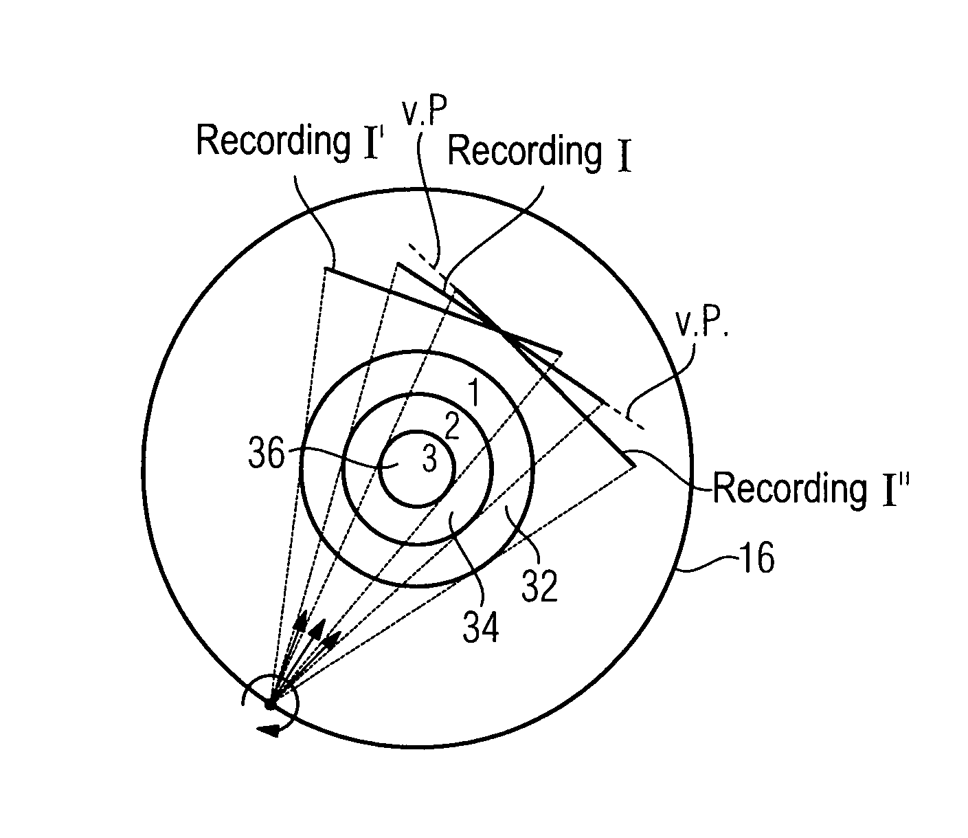
[0022]FIGS. 2 and 3 show a circular arc 16 defined by a fall rotation of an x-ray image recording system comprising x-ray source and x-ray detector, e.g. by a 360-degree rotation of an x-ray C-arm. A marked point 18 in each case indicates the origin of x-ray radiation, in other words the focus within an x-ray source. The point 18 is simultaneously a center of rotation for the x-ray source, the circular arrow 20 illustrating the rotation. The x-ray radiation is radiated in the direction of the arrows 22 in three different recordings I′, I, I″. The calibration phantom 10 from FIG. 1 is mapped if the maximum size for this is chosen. In this case the expansion of the x-ray radiation emitted by the x-ray source is not sufficiently large that all parts of the calibration phantom 10 are mapped on recording I′, the first different projection image. Nor is the calibration phantom 10 fully visible on recording I″, the second different projection image. It is now a matter of creating a virtual...


PUM


Abstract

The invention relates to a method for determining gray-scale values for volume elements of bodies to be mapped using an x-ray image recording system. When a body to be mapped is not mapped in full on a single projection image for a rotational position, a second projection image must be made and a virtual projection image derived from the two projection images, this being back-projected onto the volume elements. For calibration the present invention proposes making the same two projection images in each case at a calibration phantom and additionally a further projection image, corresponding to the position and orientation of the virtual projection image. As a result the mathematical relationships between the projection images and the virtual projection image and for the back-projection can be derived.

Description

CROSS REFERENCE TO RELATED APPLICATIONS[0001]This application claims priority of German application No. 10 2007 003 877.3 filed Jan. 25, 2007, which is incorporated by reference herein in its entirety.FIELD OF THE INVENTION[0002]The invention relates to a method for determining gray-scale values for volume elements of bodies to be mapped using an x-ray image recording system having an x-ray source and x-ray detector.BACKGROUND OF THE INVENTION[0003]The method emerges directly from the method for reconstruction of a three-dimensional image volume described in DE 10 2006 041 033.5 published after the filing date of the present application. Whereas with conventional methods for determining gray-scale values for volume elements of bodies to be mapped at predetermined rotational positions of the x-ray image recording system a 2D x-ray image (projection image) is recorded in each case, DE 10 2006 041 033.5 deals with the problem that this projection image is not sufficient to fully map a ...

Claims


Application Information

Patent Timeline
Patent Type & Authority Patents(United States)
IPC IPC(8): G01D18/00
CPCA61B6/032A61B6/4028A61B6/583
Inventor HEIGL, BENNOHOPPE, STEFANHORNEGGER, JOACHIMLAURITSCH, GUNTER
Owner SIEMENS HEALTHCARE GMBH
Why Eureka
Try Eureka