Ultrasonic image processing apparatus and control program for ultrasonic image processing apparatus

a technology of ultrasonic image processing and control program, which is applied in the direction of ultrasonic/sonic/infrasonic diagnostics, applications, instruments, etc., can solve the problems of many peaks of myocardial velocity, inability to accurately determine whether, and heart failur

Inactive Publication Date: 2010-11-02
TOSHIBA MEDICAL SYST CORP
View PDF15 Cites 44 Cited by
  • Summary
  • Abstract
  • Description
  • Claims
  • Application Information

AI Technical Summary

Benefits of technology

[0013]The present invention has been made in consideration of the above problems, and has as its object to provide a medical image processing apparatus which allows correct comprehension of the motion state of a tissue in a subject to be examined, and a control program for the medical image processing apparatus.

Problems solved by technology

As a result, dyssynchrony occurs in the contraction of the cardiac wall, and blood cannot be sufficiently pumped out, resulting in heart failure.
This is because it cannot be accurately determined whether the cause of a heart failure case is systolic dyssynchrony.
The following problems, however, arise in the above conventional techniques.
In a heart failure case, in particular, it is known that the myocardial velocity has many peaks, because the cardiac muscle moves abnormally.
That is, it is difficult to perform stable evaluation from a motion velocity to determine which peak is a peak indicating significant ventricular contraction.
According to the technique disclosed in patent reference 1, therefore, since the possibility of erroneously detecting a peak indicating significant contraction in a heart failure case is high, it is difficult to reliably detect systolic dyssynchrony.

Method used


Image

  • Ultrasonic image processing apparatus and control program for ultrasonic image processing apparatus
  • Ultrasonic image processing apparatus and control program for ultrasonic image processing apparatus
  • Ultrasonic image processing apparatus and control program for ultrasonic image processing apparatus

Examples


Embodiment Construction

[0029]An embodiment of the present invention will be described below. This embodiment will exemplify a case wherein the local motion of a cardiac muscle tissue of the heart as a target is evaluated by using a two-dimensional image.

(Arrangement)

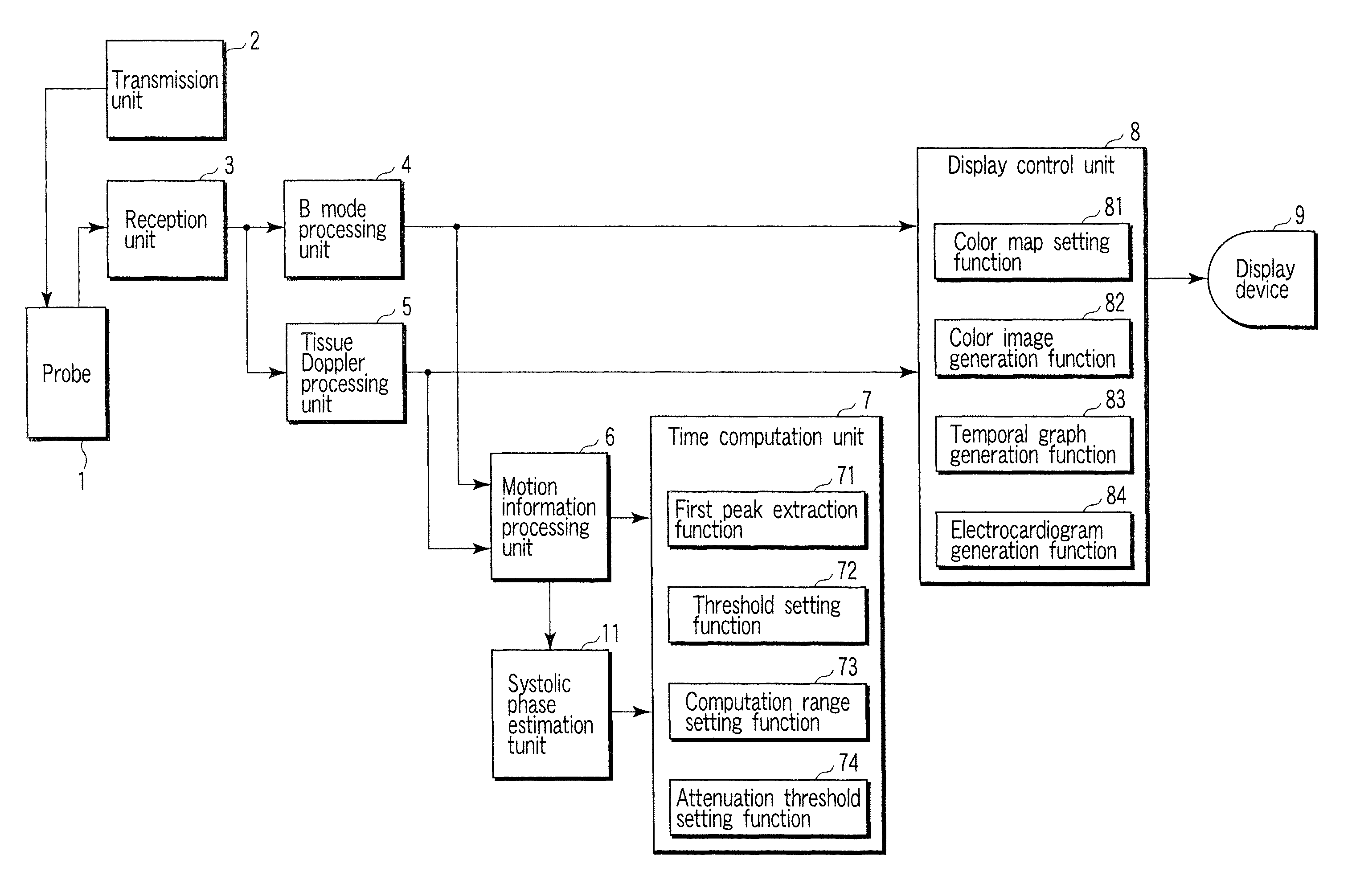
[0030]FIG. 1 is a block diagram showing the arrangement of an ultrasonic image processing apparatus according to an embodiment of the present invention. An ultrasonic probe 1 includes an ultrasonic transducer array of a plurality of ultrasonic transducers which convert electrical signals into ultrasonic waves. Ultrasonic waves are transmitted / received to / from a subject by using this ultrasonic transducer array. Assume that in the first embodiment, the ultrasonic probe 1 is a sector probe targeted to the heart.

[0031]A transmission unit 2 generates driving signals for transmitting ultrasonic waves from the ultrasonic transducer array. The transmission unit 2 generates a driving signal having a predetermined delay characteristic for each transduc...


PUM


Abstract

Motion information of a tissue at a plurality of positions in a subject to be examined which is obtained by transmission / reception of an ultrasonic wave is acquired. This information includes, for example, a quantitative value associated with the strain of the tissue in the subject or a quantitative value associated with a displacement in the subject. A predetermined motion timing of the subject is calculated on the basis of the motion information of the tissue. A first image for indicating the motion timings of the tissue at a plurality of positions in the subject is generated.

Description

CROSS-REFERENCE TO RELATED APPLICATIONS[0001]This application is based upon and claims the benefit of priority from prior Japanese Patent Application No. 2005-376261, filed Dec. 27, 2005, the entire contents of which are incorporated herein by reference.BACKGROUND OF THE INVENTION[0002]1. Field of the Invention[0003]The present invention relates to an ultrasonic image processing apparatus which generates an image indicating the motion information of a subject to be examined on the basis of echo information obtained by transmitting / receiving ultrasonic waves to / from the subject, and a control program for the ultrasonic image processing apparatus.[0004]2. Description of the Related Art[0005]There is known an ultrasonic diagnostic apparatus which scans the interior of a subject with ultrasonic waves and visualizes the interior of the subject on the basis of reception signals generated from reflected waves from the interior of the subject. This ultrasonic diagnostic apparatus transmits ...

Claims


Application Information

Patent Timeline
Patent Type & Authority Patents(United States)
IPC IPC(8): A61B8/00G06T1/00G06T7/20
CPCA61B8/463A61B8/485A61B8/488G01S7/52042G01S7/52071G01S7/52087G01S15/8979A61B8/0883G01S7/52074
Inventor ABE, YASUHIKOKAWAGISHI, TETSUYA
Owner TOSHIBA MEDICAL SYST CORP
Why Eureka
Try Eureka