Image evaluation method for two-dimensional projection images and objects corresponding thereto

a two-dimensional projection and image evaluation technology, applied in the field of image evaluation methods for two-dimensional projection images, can solve the problems of complex classifications, significant risk of artifacts, and difficulty in determining the quantity of blood supply to the myocardium through angiographic methods

Inactive Publication Date: 2011-01-04
SIEMENS HEALTHCARE GMBH
View PDF12 Cites 2 Cited by
  • Summary
  • Abstract
  • Description
  • Claims
  • Application Information

AI Technical Summary

Benefits of technology

[0127]It is possible for the computer to retain the sub-parcels as such. However it is preferable for the computer to combine sub-parcels, to which it does not assign the type “vessel” in the new iteration, with a parcel adjacent to the relevant sub-parcel, to which the type “vessel” is also not assigned, to form an overall parcel. This measure can reduce computation outlay.
[0129]It is possible for the computer to change the type to “background” for parcels, to which it assigned the type “perfusion area”, if the relevant parcel is surrounded in an angle range, which is larger than a second minimum angle range, by parcels, to which the computer has assigned the type “background”. It is possible in particular to eliminate outliers with this procedure.
[0132]It is possible for the computer to carry out a vessel segmentation for each projection image based on the pixel values of the respective projection image and to take into account the vessels identified based on the vessel segmentation during the type assignment. This procedure allows more defined separation of the vessels of the vascular system from the remainder of the image.
[0134]The evaluation of the projection images is on the one hand particularly simple and on the other hand particularly true to reality, if the computer uses the parameters to determine a characteristic input time for inputting the contrast medium into the corresponding evaluation core for a pixel under consideration and a characteristic washout time for washing the contrast medium out from the corresponding evaluation core and determines the degree of perfusion based on the input and washout times.

Problems solved by technology

Quantification of the supply of blood to the myocardium by means of angiographic methods is however problematic, since the angiographically observable cardiac vessels have a diameter of just under a millimeter or more.
The classifications are complex and can be applied only in a time-consuming manner.
This gives rise to a significant risk of artifacts, to avoid which computation-intensive methods are in turn required in order to compensate for motion.

Method used


Image

  • Image evaluation method for two-dimensional projection images and objects corresponding thereto
  • Image evaluation method for two-dimensional projection images and objects corresponding thereto
  • Image evaluation method for two-dimensional projection images and objects corresponding thereto

Examples


Embodiment Construction

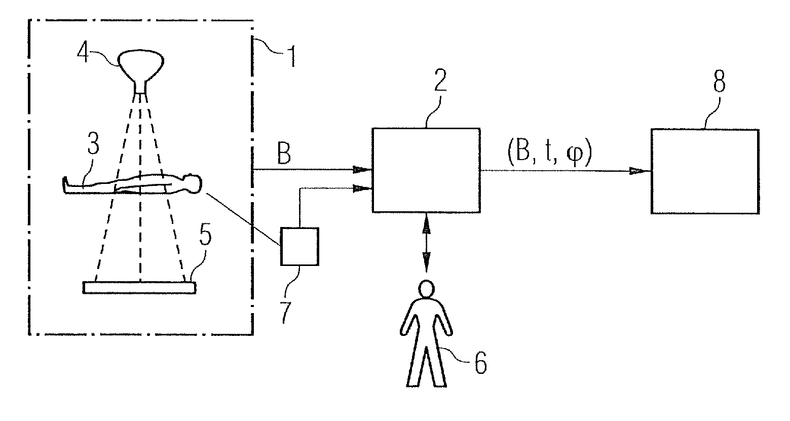
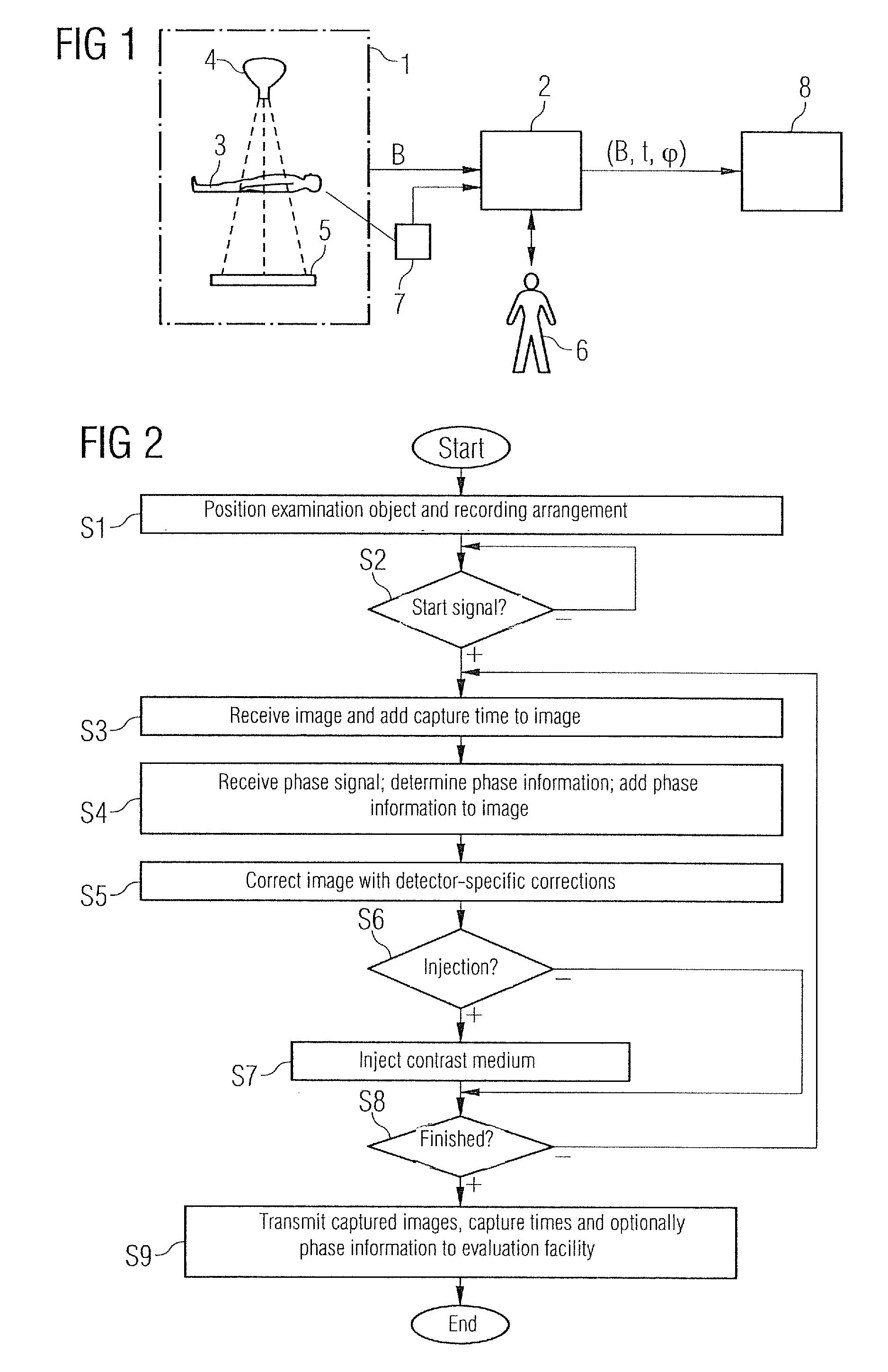
[0162]Large sections of the present invention correspond to the procedure described in DE 10 2005 039 189.3. The above comments relating to FIGS. 1 to 8 also apply within the scope of the present invention. Thus for example the present invention relates not only to an image evaluation method as such but also—see also FIG. 4—to a data medium 13 with a computer program 12 stored on the data medium 13, to carry out an inventive image evaluation method. It also relates—see also FIG. 4 again—in addition to the image evaluation method to a computer 8 with a mass memory 11, in which a computer program 12 is filed, so that the computer 12 executes an evaluation method of this type after the computer program 12 has been called.

[0163]The essential difference between the present invention and the procedure in DE 10 2005 039 189.3 is the implementation of steps S35 to S37 in FIG. 6. These differences are examined in more detail below. The wording of DE 10 2005 039 189.3 and the reference charac...


PUM


Abstract

2-D projection images show the temporal profile of the distribution of a contrast medium in an examination object, which contains a vascular system and its surroundings. Each projection image comprises pixels with pixel values. The pixel values of pixels corresponding to one another in the projection images are defined by at least essentially locationally identical areas of the examination object. A computer assigns a uniform 2-D evaluation core that is uniform for all corresponding pixels at least in a sub-area of pixels corresponding to one another in the projection images that is uniform for the projection images. The computer defines at least one characteristic value for each pixel within each projection image based on the evaluation core assigned to the pixel and assigns it to the relevant pixel. Based on the temporal profile of the characteristic values, the computer defines parameters of at least one function of time, so that any deviation between the function parameterized with the parameters and the temporal profile of the characteristic values is minimized. Based on the parameters the computer defines a type and / or an extent and assigns them to a pixel of a 2-D evaluation image corresponding to the pixels of the projection images. The type indicates whether the respective pixel of the evaluation image corresponds to a vessel of the vascular system, a perfused part or a non-perfused part of the surroundings of a vessel of the vascular system. The extent is characteristic of perfusion. The computer outputs at least the sub-area of the evaluation image to a user via a display device.

Description

CROSS REFERENCE TO RELATED APPLICATIONS[0001]This application claims priority of German application No. 10 2006 025 422.8 DE filed May 31, 2006, which is incorporated by reference herein in its entirety.FIELD OF INVENTION[0002]The present invention relates to an image evaluation method for two-dimensional projection images, which show the temporal profile of the distribution of a contrast medium in an examination object, the examination object containing a vascular system and its surroundings, each projection image comprising a plurality of pixels having pixel values, the pixel values of pixels corresponding to one another in the projection images being defined by at least essentially locationally identical areas of the examination object.[0003]The present invention relates furthermore to a data medium with a computer program stored on the data medium for implementing an image evaluation method of this type and to a computer with a mass memory in which a computer program is filed, s...

Claims


Application Information

Patent Timeline
Patent Type & Authority Patents(United States)
IPC IPC(8): G06K9/00A61B5/02
CPCG06T7/0012A61B6/507G06T2207/30104G06T2207/10116
Inventor CAMUS, ESTELLEREDEL, THOMAS
Owner SIEMENS HEALTHCARE GMBH
Why Eureka
Try Eureka