Solid phase isolation of proteins, nucleic acids and other macromolecules

a technology of protein and nucleic acid, applied in the field of solid phase isolation of proteins, nucleic acids and other macromolecules, can solve the problems of difficult to remove the molecule of interest from the microtiter plate, polystyrene is not stable, and the isolation of a sufficient quantity of a molecule is not typically allowed to perform further tests

Inactive Publication Date: 2011-07-19
CA RGT UNIV OF
View PDF16 Cites 3 Cited by
  • Summary
  • Abstract
  • Description
  • Claims
  • Application Information

AI Technical Summary

Benefits of technology

[0008]The invention is a method for isolating macromolecules comprising coating the inner walls of a test tube with beads to a predetermined level; coating the beads with a capture reagent of the macromolecule of interest; incubating the coated beads with a solution containing the macromolecule under conditions to allow binding of the macromolecule to the capture reagent; washing the coated beads with the bound macromolecule with a wash buffer to remove unbound material while maintaining binding of the macromolecule to the binding partner; and eluting the macromolecule from the binding partner. This method allows for affinity purification of macromolecules in a substantially more rapid fashion than more typical immunoprecipitation methods, column chromatography or other methods in which a capture reagent attached to beads for isolation of a macromolecule of interest wherein the beads are not attached to a solid support.

Problems solved by technology

Such assays allow for the determination of the presence of a molecule in a sample, but do not typically allow for the isolation of a sufficient quantity of a molecule to perform further tests.
Moreover, it is difficult to remove the molecule of interest from microtiter plates.
Polystyrene is not stable in the presence of a variety of organic compounds frequently used for elution.
Additionally, the plates cannot be heated to elute the samples into loading buffer for analysis of the samples by SDS-polyacrylamide gel electrophoresis (PAGE).
The use of these techniques is limited both by the cost of the reagents required (e.g. protein A- or protein G-agarose) and the amount of effort required to perform the assays.
This limits the number of samples that can be processed by an individual at one time and makes automation of the process difficult.
Due to the number of washes required, it is not practical to process more samples than can be placed in a single microfuge rotor, typically 18.
), which is both uncomfortable and impractical.
Although such methods are somewhat more amenable to automation, elution of the molecule of interest from the column requires a relatively large amount of buffer, frequently resulting in a solution of the molecule of interest that must be concentrated before further analysis.
This is time consuming and can result in deterioration of the sample.

Method used


Image

  • Solid phase isolation of proteins, nucleic acids and other macromolecules
  • Solid phase isolation of proteins, nucleic acids and other macromolecules
  • Solid phase isolation of proteins, nucleic acids and other macromolecules

Examples


example 1

[0059]Bonding of glass beads to the inner walls of microcentrifuge tubes. Acid-washed glass microbeads, 425-600 μm diameter, were obtained from Sigma-Aldrich Co.; 3-aminopropyltriethoxysilane and dimethyl suberimidate were from Pierce Chemical Co.; and conical polypropylene microcentrifuge tubes [12.6 mm (O.D.)×47.6 mm (L), 1.5 ml volume, screw-cap with O-ring] were from Fisher Scientific.

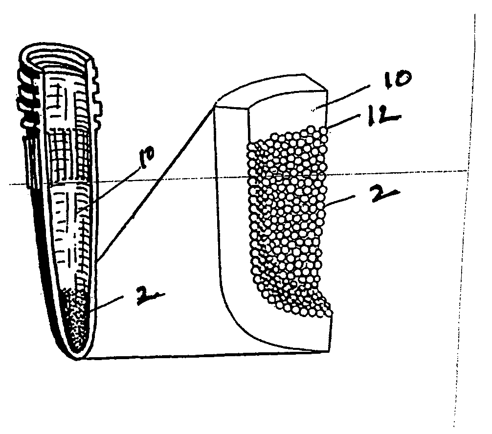
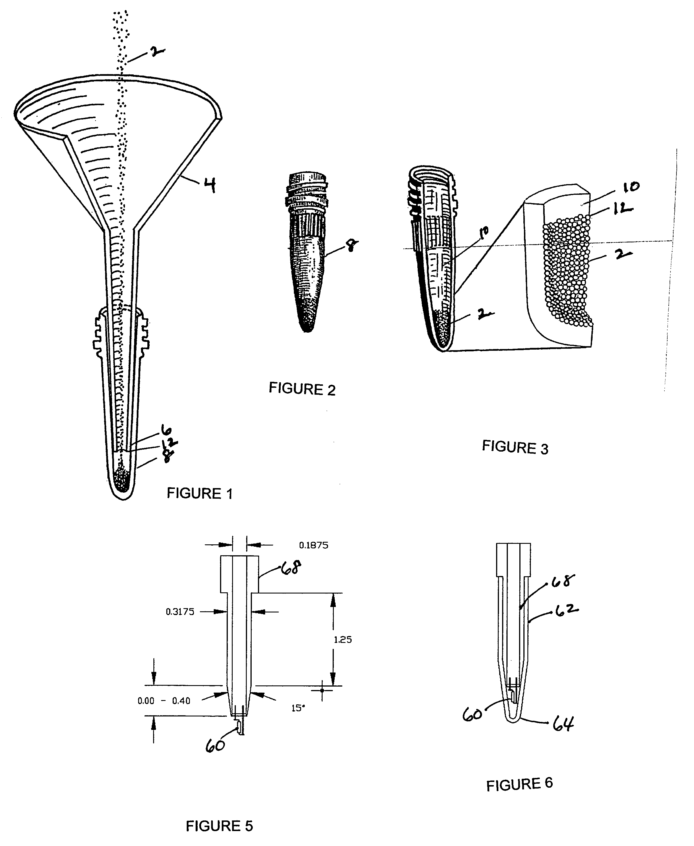
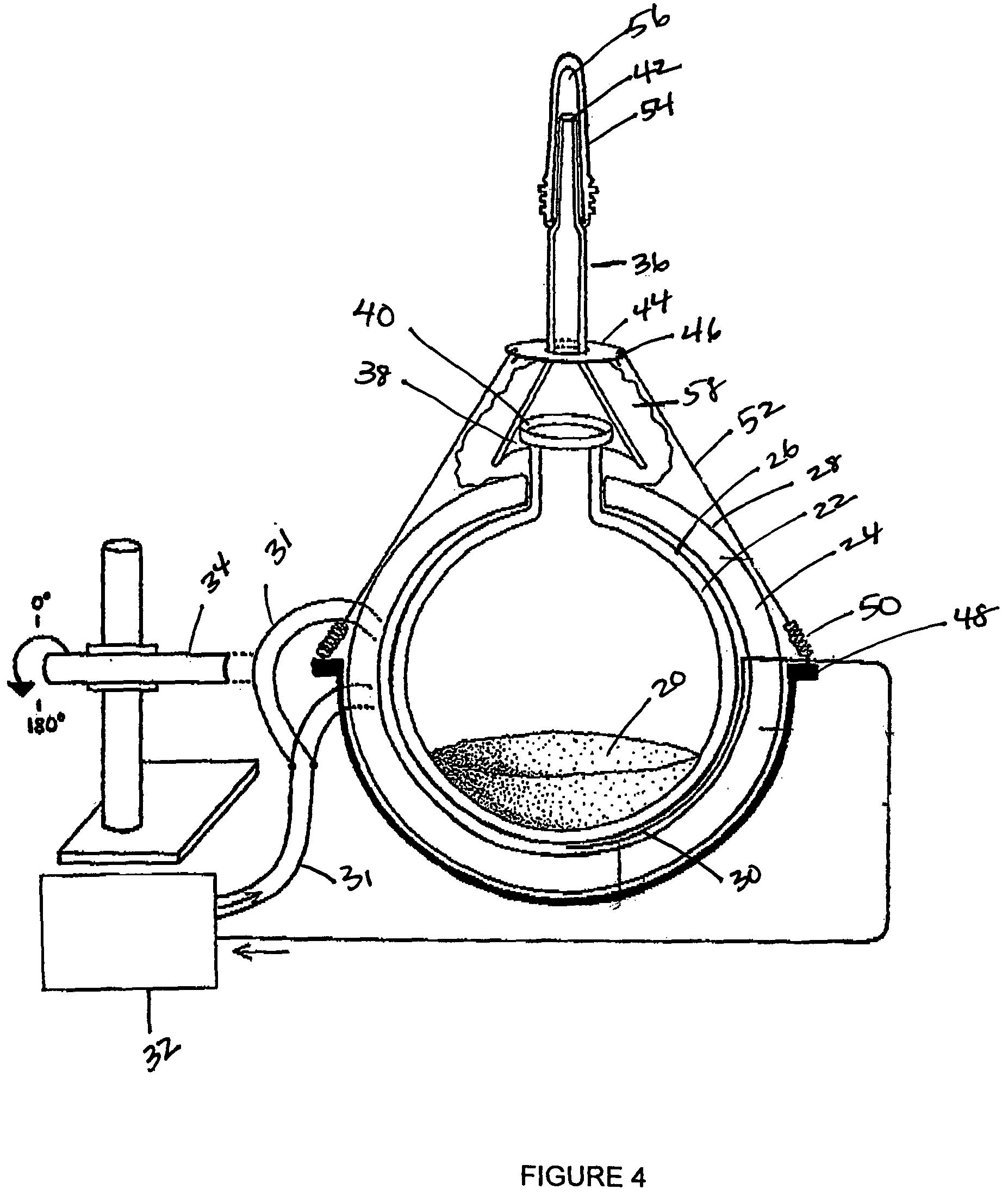
[0060]Glass microbeads were heated to 455° C. in a round bottom flask by upper and lower heating mantles under control of a microprocessor-controlled temperature regulator. The heated beads were added to microcentrifuge tubes by pouring through a glass funnel inserted into the tubes (see FIG. 1). The high temperature of the beads melted the superficial layer of the inner wall of the tubes, and the beads became bonded to the tube wall; excess beads which did not bond to the tubes were returned to the round bottom flask. Within the tapered portion of the microcentrifuge tubes, the height of the beads...

example 2

[0063]Immobilization of protein A or protein G on glass Microbeads. After bonding the glass microbeads to the wall of the centrifuge tubes, protein A or G was immobilized on the glass beads. To accomplish this, free Si groups on the bead surface were first reacted with aminopropyltriethoxysilane to yield free amino groups; aminopropyltriethoxysilane diluted 1:5 in acetone was incubated at room temperature for 5 min with the bonded beads, and the beads were washed three times with acetone and three times with 100 mM Hepes, pH 7.4. The homobifunctional imidoester dimethyl suberimidate was then used to cross-link the free amino groups on the surface of the glass beads to amino groups of protein A or G. This reaction proceeded for 1 h at 4° C. using 1 mM dimethyl suberimidate and 1 mg / ml of protein A or G in 100 mM Hepes, pH 8.5. Under these conditions, the preferred reaction is with the α-amines of the proteins rather than with epsilon amines of glutamine and asparagine. The reaction w...

example 3

[0064]Quantitation of protein A binding sites on glass microbeads. The number of protein A binding sites on the glass beads bonded to the wall of microcentrifuge tubes was determined by two different methods. In the first method, 50 μg of rabbit IgG in HNMN buffer (50 mM Hepes, pH 7.5, 150 mM NaCl, 5 mM MgCl2, 1% Nonidet P-40) was added to the tubes, and the tubes were shaken gently for 2 h at 4° C. to allow for binding of the rabbit IgG to the immobilized protein A. The beads were washed three times with Tris buffer and IgG bound to the beads was measured by the Bradford method using bovine serum albumin as a standard.

[0065]In the second method, 2-10 μg of 125I-labeled rabbit anti-mouse IgG (specific activity 10 μCi / pg) was added to the tubes, and after a 1 hour incubation and six washes, the amount of immunoglobulin bound to the beads was determined by gamma counting and comparison to a standard curve.

[0066]Upon analysis of total protein bound to the glass beads, it was determined...


PUM

PropertyMeasurementUnit
melting pointaaaaaaaaaa
melting temperatureaaaaaaaaaa
diameteraaaaaaaaaa

Abstract

The invention is a method for the isolation of molecules of interest using tubes in which at least a portion of the inner walls of the tube are coated with microbeads that are coated with a capture reagent to bind the molecule of interest. The microbeads may be glass or polymer beads. The invention is a method and apparatus for preparation of the tubes for use in the method of the invention. The invention is a method for determining ratios of guanine nucleotides bound to guanine-nucleotide binding proteins.

Description

CROSS REFERENCE TO RELATED APPLICATIONS[0001]This application claims the benefit of priority of U.S. provisional application Ser. No. 60 / 443,231 filed Jan. 28, 2003 which is incorporated herein by reference in its entirety.GOVERNMENT INTEREST[0002]This invention was made with government support under CA76968 awarded by the National Institutes of Health. The government has certain rights in the invention.BACKGROUND OF THE INVENTION[0003]Solid-phase capture and affinity purification have a wide variety of applications in research, clinical, and pharmaceutical laboratories. For the batch analysis of multiple samples, a capture reagent (e.g., an antibody) can be immobilized on the bottom of microtiter plate wells to bind a molecule of interest (e.g., the corresponding antigen). This system provides a relatively small surface area for capture and a small working volume during binding and washing of the target molecule. Such assays allow for the determination of the presence of a molecule...

Claims


Application Information

Patent Timeline
Patent Type & Authority Patents(United States)
IPC IPC(8): G01N33/543C07K1/22C07K14/47
CPCC07K1/22C07K14/4722
Inventor BOSS, GERRY R.JONES, STEPHEN B.CHEN, JEFFREY C.VON LINTIG, FRIEDERIKEHOFFMAN, STEPHANIEKIMMERLY, ADAMLUU, JOANNMYCROFT, MATTHEW CHRISTOPHER
Owner CA RGT UNIV OF
Why Eureka
Try Eureka