Esophageal mapping catheter

a technology of esophageal mapping and catheter, which is applied in the field of esophageal mapping catheter, can solve the problems of high mortality rate, esophageal fistula formation, and damage to the esophagus, and achieve the effects of reducing the risk of energy delivery, and reducing the risk of esophageal fistula formation

Active Publication Date: 2012-07-17
BIOSENSE WEBSTER INC
View PDF20 Cites 68 Cited by
  • Summary
  • Abstract
  • Description
  • Claims
  • Application Information

AI Technical Summary

Benefits of technology

[0008]The present invention generally relates to a catheter-based solution to the above-described problem that provides the user the ability to record and display the location of the esophagus on an electroanatomic mapping system such as the Biosense Webster CARTO™ System. Esophagus location information will enable the user to perform RF ablations in the left atrium such that the esophagus is not in close proximity to the ablation sites, reducing the risk of energy delivery close to the esophagus, thus reducing the risk of esophageal fistula formation.
[0009]The esophageal mapping catheter of the current invention includes a flexible, tubular device with a location sensor and thermocouples located in the device tip. The device is introduced into the esophagus via the throat or nasal passage and is aligned behind the heart using fluoroscopic guidance. Prior to performing left atrial (LA) ablations, the physician records location data points within the esophagus by advancing and withdrawing the device within the esophagus while recording catheter location information from the location sensor, tagging location points. The points serve to record device tip position within the esophagus, in turn recording the relative position of the esophagus with respect to the left atrium on the electroanatomic map. Esophagus points displayed on the electroanatomic map enable the physician to direct RF energy delivery away from the esophagus reducing the risk of esophageal fistula formation.
[0010]In addition, the device can be placed within the esophagus, behind the left atrium, during RF energy delivery to measure esophagus temperatures. Esophagus temperature changes during ablation may provide the user feedback during the ablation to prevent thermal damage to the esophagus.
[0011]The catheter placement in the esophagus can be guided using x-ray to insure that the radiopaque catheter aligns along the posterior wall of the left atrium. The device may be coated with a lubricious material to facilitate easy introduction into and manipulation within the esophagus.
[0012]The present invention is a flexible device, suitable for introduction into the esophagus that is instrumented to allow for recording of esophagus position with respect to the ablation site during an electrophysiology ablation procedure. Esophagus position information enables user to direct ablation away from the esophagus reducing risk of esophageal fistula formation. Temperature information may allow the user to control power delivery during ablation to prevent thermal damage to the esophagus.

Problems solved by technology

Some procedures require the use of the catheter-delivered energy sources near the esophagus resulting in the risk of damage to the esophagus including the creation of esophageal fistula.
Esophageal fistula associated with ablation used to treat atrial fibrillation have resulted in a high incidence of mortality.

Method used


Image

  • Esophageal mapping catheter
  • Esophageal mapping catheter
  • Esophageal mapping catheter

Examples


Embodiment Construction

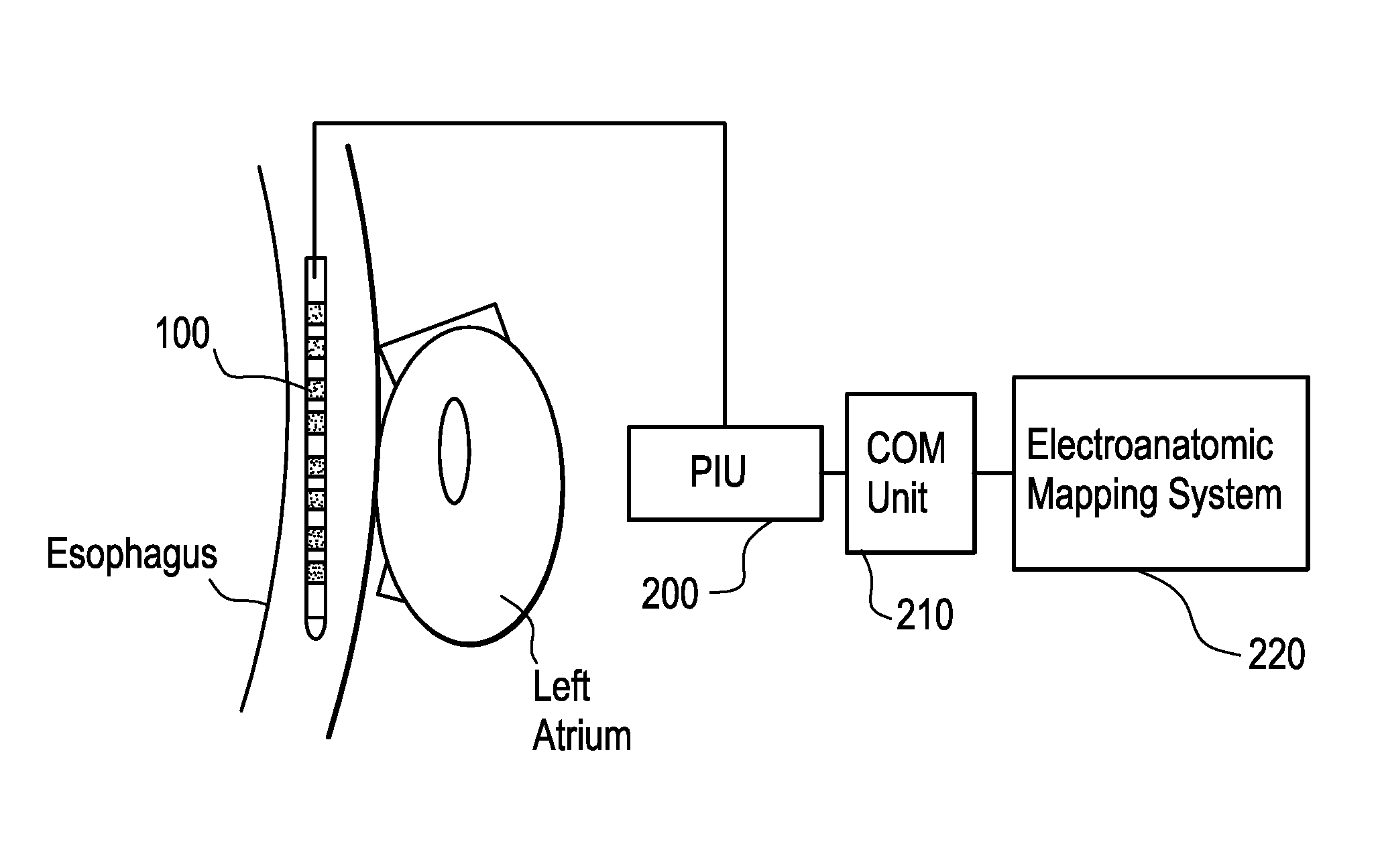
[0023]FIG. 1 is a diagram depicting the placement of an esophageal mapping catheter 100 in the esophagus near the left atrium of the heart of a patient. Esophageal mapping catheter 100 is electrically connected to the patient interface unit (PIU) 200 in communication with the communications (COM) unit 210 which is further in communication with the electroanatomic mapping system 220. Electrical signals from the esophageal mapping catheter 100 are thereby received and operated upon by the electroanatomic mapping system 220 as described below.

[0024]The close anatomical relationship of the posterior wall of the left atrium of the heart and the thermosensitive esophagus, creates a potential hazard in catheter ablation procedures. Esophageal mapping catheter 100 is introduced through the patient's nose or throat into the esophagus. Once in the desired position, the device's location sensor is used to “tag” the 3-D position of the esophagus lumen, using the location software executed in th...


PUM


Abstract

An esophageal mapping catheter enables a physician to map the location of the esophagus so as to avoid damaging the esophagus during radio frequency (RF) ablation procedures. Information from the esophageal mapping catheter is communicated to a patient information unit, communications unit and / or electroanatomic mapping system. The electroanatomic mapping system uses the information from the esophageal mapping catheter to develop a three-dimensional map of the esophagus and to monitor the temperature within the esophagus in order to prevent the creation of esophageal fistula.

Description

CROSS REFERENCE TO RELATED APPLICATION[0001]This application claims the benefit of U.S. Provisional Application No. 60 / 828,885, filed Oct. 10, 2006, which is incorporated by reference herein.FIELD OF THE INVENTION[0002]The present invention relates to a device for catheter-based anatomic mapping of the esophagus for use during an electrophysiology ablation procedure. More particularly, the device is placed in the esophagus via the transpharyngeal or transnasal approach to provide esophagus reference location and orientation data during an ablation procedure.BACKGROUND OF THE INVENTION[0003]Electrophysiology ablation procedures use energy sources, such as radio frequency (RF) energy, to ablate tissue in order to correct or prevent arrhythmias such as super ventricular tachycardia, paroxysmal atrial tachycardia or atrial fibrillation. Some procedures require the use of the catheter-delivered energy sources near the esophagus resulting in the risk of damage to the esophagus including t...

Claims


Application Information

Patent Timeline
Patent Type & Authority Patents(United States)
IPC IPC(8): A61B5/05A61B5/296
CPCA61B1/0008A61B5/4233A61B5/06A61B1/2733A61B5/062A61B1/00097
Inventor MOTTOLA, BRIANO'SULLIVAN, MARTIN F.WU, SUE-LYNN
Owner BIOSENSE WEBSTER INC
Why Eureka
Try Eureka