
Orthopedic procedures training simulator

a training simulator and orthopaedic technology, applied in the field of orthopaedic procedures training simulators, can solve the problems of only limited experience in performing orthopaedic procedures on actual patients, difficult to learn orthopaedic procedures for medical students, and further injury to patients

Inactive Publication Date: 2013-05-14
WILKINS JASON D
View PDF26 Cites 9 Cited by
  • Summary
  • Abstract
  • Description
  • Claims
  • Application Information

AI Technical Summary

Benefits of technology

[0010]Larger joints, such as the shoulder and hip joints, employ a system of motor-tensioned cables to join adjacent skeletal members. Cables may be located about each joint to simulate muscles and tendons of the human anatomy, providing for realistic mechanical action and feel of the joints. A computer system controls the motors to tension or relax the cables, allowing the shoulder and hip joints to be selectively dislocated or reduced. The computer system accepts entry of medication orders, including sedative, local or regional anesthetic, and other medications, to vary the tension applied to the cables, allowing procedural training to include use of medication, such as a sedative, to facilitate reduction of a dislocated shoulder or hip.
[0014]Thus, the orthopedic procedures training simulator allows a student or trainee unlimited opportunity for training and practice of orthopedic procedures, including the administration and effect of various medications, without the risks attendant to performing the procedures on an actual patient.

Problems solved by technology

Orthopedic procedures are difficult for medical students to learn, largely because of limited opportunities for students and residents to perform the procedures.
Medical students and residents are allowed only limited experiences in performing orthopedic procedures on actual patients, both because of limited availability of patients with certain injuries requiring specialized procedures and because of the risk that procedural errors by an untrained student or resident might lead to further injury to the patient.
While the anatomical and structural insights that may be gained by use of a medical model may inform a student on the function and need for a particular procedure to correct a given injury, such models are generally limited in scope and are functionally inadequate in actually simulating a given injury to allow practice of a corrective orthopedic procedure.
While some medical models have been devised to illustrate orthopedic injuries, and in some devices even allowing demonstration of a mechanical reduction of the injury, simple mechanical aspects of an injury and reduction procedure are often insufficient for adequate training.
Because diagnosis is typically made based on x-ray images along with external observation and palpation, certain medical models, such as skeletal models, are inappropriate for diagnostic training.
A simple mechanical model lacks the ability to first, simulate a condition suggesting the need for such medication, and second, simulate a physical response to the administration of the medication.

Method used


Image

  • Orthopedic procedures training simulator
  • Orthopedic procedures training simulator
  • Orthopedic procedures training simulator

Examples


Embodiment Construction

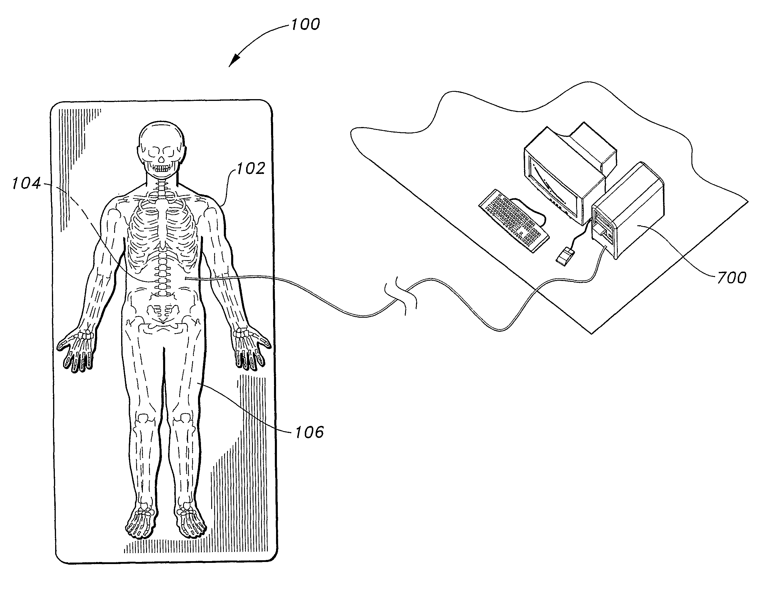
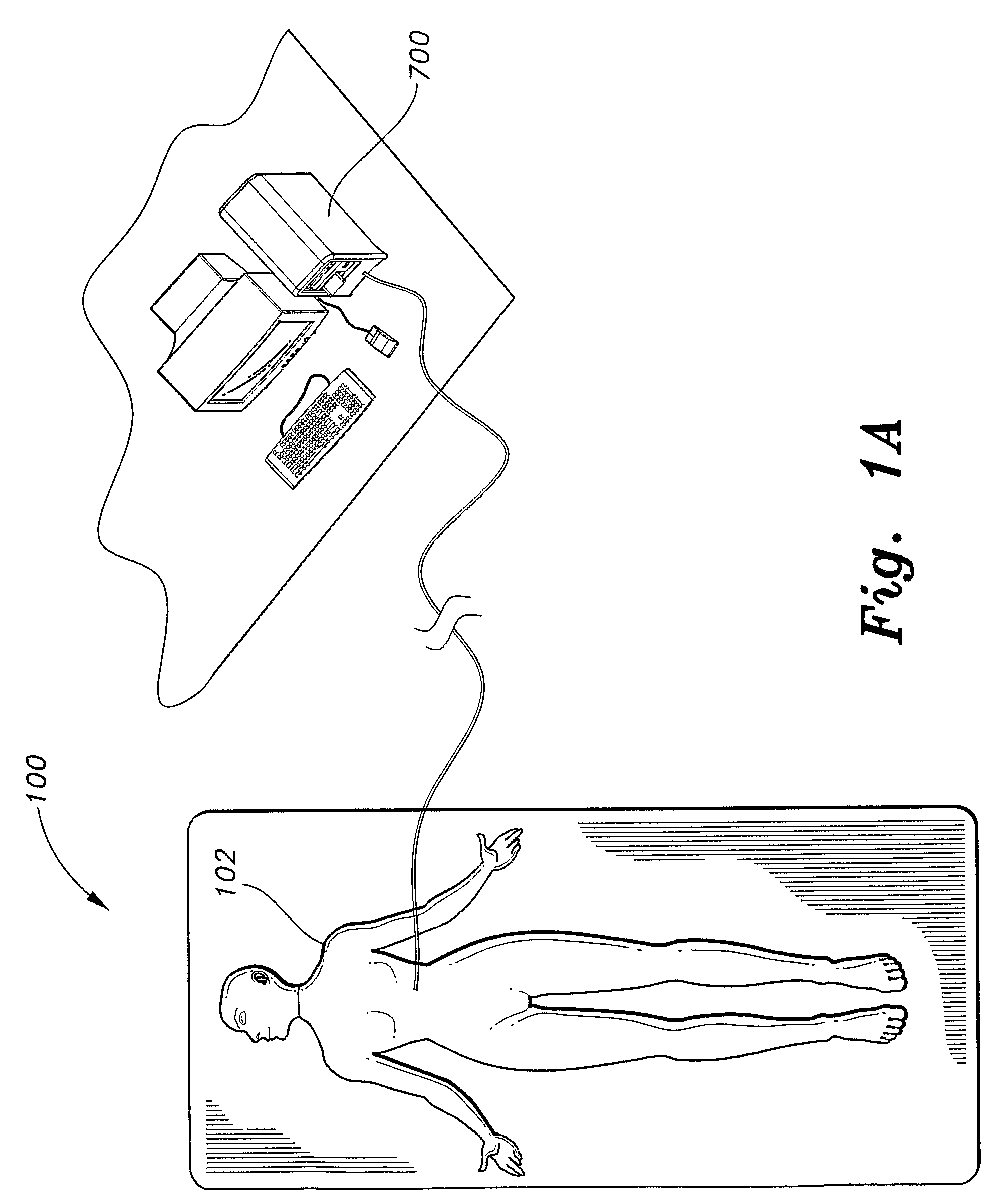
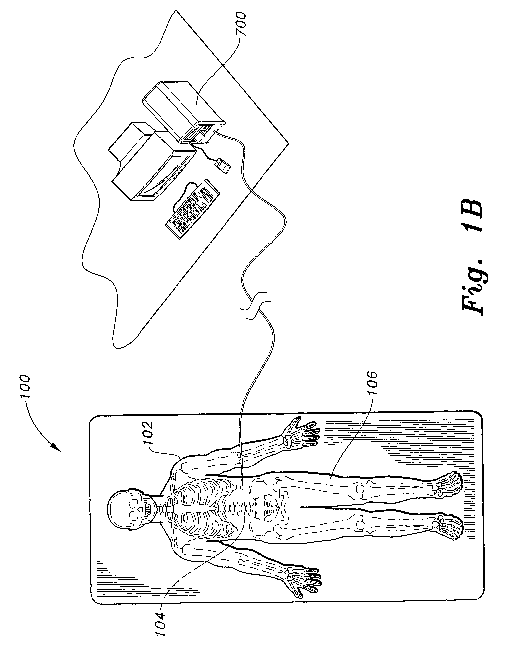
[0034]The present invention is an orthopedic procedures training simulator, designated generally as 100 in the figures. Referring to FIGS. 1A-1C, the orthopedic procedures training simulator 100 is a figure or mannequin 102 generally, in the illustrated embodiment, in the form of a human body having an internal skeletal structure 104 adapted for simulating various orthopedic noxious events including joint dislocations and bone fractures.

[0035]The internal skeletal structure 104 is comprised of numerous skeletal members arranged generally in the form of a human skeleton. A covering 106 of foam or similar material surrounds the internal skeletal structure 104 to give the mannequin 102 a somewhat realistic appearance of a human body.

[0036]The internal skeletal structure 104 is instrumented by stress and torque sensors, position sensors, and electrical contacts located on and near various skeletal members of the internal skeletal structure 104. A computer system 700 is in communication ...


PUM


Abstract

The orthopedic procedures training simulator (100) is a mannequin (102) having an internal skeletal structure (104) for simulating orthopedic injuries. The interior skeletal structure (104) includes a plurality of skeletal structures, each movable between a first position simulating normal and healthy skeletal structure and a second position simulating an orthopedic injury. Motor tensionable cables joining larger joint structures allow for simulating muscular tension affecting the ability to reduce a dislocation of the joint. Force and positional sensors are disposed within the skeletal structures, and provide information to a computer system (700) to perform and monitor simulation. A computer program reads the sensors, and controls the motor tensionable cables to simulate orthopedic injuries and responses to applied corrective procedures and medications. The program generates pre-procedural x-ray images based on a desired simulation, and post-procedural x-ray images based on information obtained from the sensors both during and after the performance of a procedure.

Description

CROSS-REFERENCE TO RELATED APPLICATION[0001]This application is a 371 of PCT / US06 / 13259, filed Apr. 7, 2006, which claims the benefit of U.S. Provisional Patent Application Ser. No. 60 / 669,027, filed Apr. 7, 2005.TECHNICAL FIELD[0002]The present invention relates to medical training methods and devices, and more particularly to an orthopedic procedures training simulator that provides training for the diagnosis and treatment procedures of noxious orthopedic events.BACKGROUND ART[0003]Orthopedic procedures are difficult for medical students to learn, largely because of limited opportunities for students and residents to perform the procedures. Medical students and residents are allowed only limited experiences in performing orthopedic procedures on actual patients, both because of limited availability of patients with certain injuries requiring specialized procedures and because of the risk that procedural errors by an untrained student or resident might lead to further injury to the...

Claims


Application Information

Patent Timeline
Patent Type & Authority Patents(United States)
IPC IPC(8): G09B23/28G09B23/32
CPCG09B23/28G09B23/30
Inventor WILKINS, JASON D.
Owner WILKINS JASON D
Learn More