Image reconstruction incorporating organ motion

a motion reconstruction and organ technology, applied in the field of reconstruction images, can solve the problems of invalid 2d motion models for imaging of the torso, inability to accurately reproduce rigid 2d motion models, etc., to achieve the effect of reducing motion artifacts, increasing the signal-to-noise ratio in the image being reconstructed, and reducing motion artifacts

Inactive Publication Date: 2012-08-09
UNIV OF UTAH RES FOUND
View PDF7 Cites 43 Cited by
  • Summary
  • Abstract
  • Description
  • Claims
  • Application Information

AI Technical Summary

Benefits of technology

[0016]The organ deformation motion can be used in reconstructing anatomical images. For example, an estimated time-indexed 4D image can then be generated using the organ deformation motion, wherein the estimated time-indexed image is viewable by an end user. The organ deformation motion estimation provided according to embodiments of the present invention can thus be used to reduce motion artifacts and / or increase the signal-to-noise ratio in images being reconstructed. Increased signal-to-noise ratio resulting from implementation of organ deformation motion estimation of embodiments facilitates a decrease in the dose of image modality energy (e.g., X-ray energy, magnetic field energy, ultrasound energy, etc.) delivered to the patient for generating an image of desired quality.

Problems solved by technology

However, at the X-ray tube currents typically used in clinical practice the signal is strong enough that the noise is approximately Gaussian.
Although application of amplitude binning algorithms in the foregoing example may reduce binning artifacts, since data is not acquired at all breathing amplitudes the images often have some missing slices.
However, such rigid 2D motion models are not valid for imaging of the torso, where respiratory-induced motion causes highly non-linear deformation with a significant component in the superior-inferior direction.
However, the acquisition of an artifact-free reference image remains impractical for many radiotherapy patients.
While the B-spline model guarantees smooth deformations, it cannot guarantee the diffeomorphic properties for large deformations and it does not directly enforce local conservation of tissue volume.
This method may be useful in reducing artifacts in a 3D image, but the optical flow model, like the B-spline model, does not ensure diffeomorphic incompressible motion estimates.
Another efficient Fourier method of incompressible projection applies a result from the continuous domain to discrete data without alteration, which does not accommodate the discrete nature of image data.
Despite these efforts in image registration, the incompressible nature of internal organs has not previously been incorporated into the image reconstruction process.
Such methods may be used with either phase or amplitude binned images, but are challenged in the presence of binning artifacts.
That is, deformable image registration is not well suited for use with respect to 4D RCCT images which include binning artifacts.
In particular, motion artifacts remain present despite application of many of the available image reconstruction techniques.
Moreover, none of the present imaging reconstruction techniques guarantee the diffeomorphic properties for large deformations or directly enforce local conservation of object volume.

Method used


Image

  • Image reconstruction incorporating organ motion
  • Image reconstruction incorporating organ motion
  • Image reconstruction incorporating organ motion

Examples


Embodiment Construction

[0033]Reference will now be made to the exemplary embodiments illustrated in the drawings, and specific language will be used herein to describe the same. It will nevertheless be understood that no limitation of the scope of the invention is thereby intended. Alterations and further modifications of the inventive features illustrated herein, and additional applications of the principles of the inventions as illustrated herein, which would occur to one skilled in the relevant art and having possession of this disclosure, are to be considered within the scope of the invention.

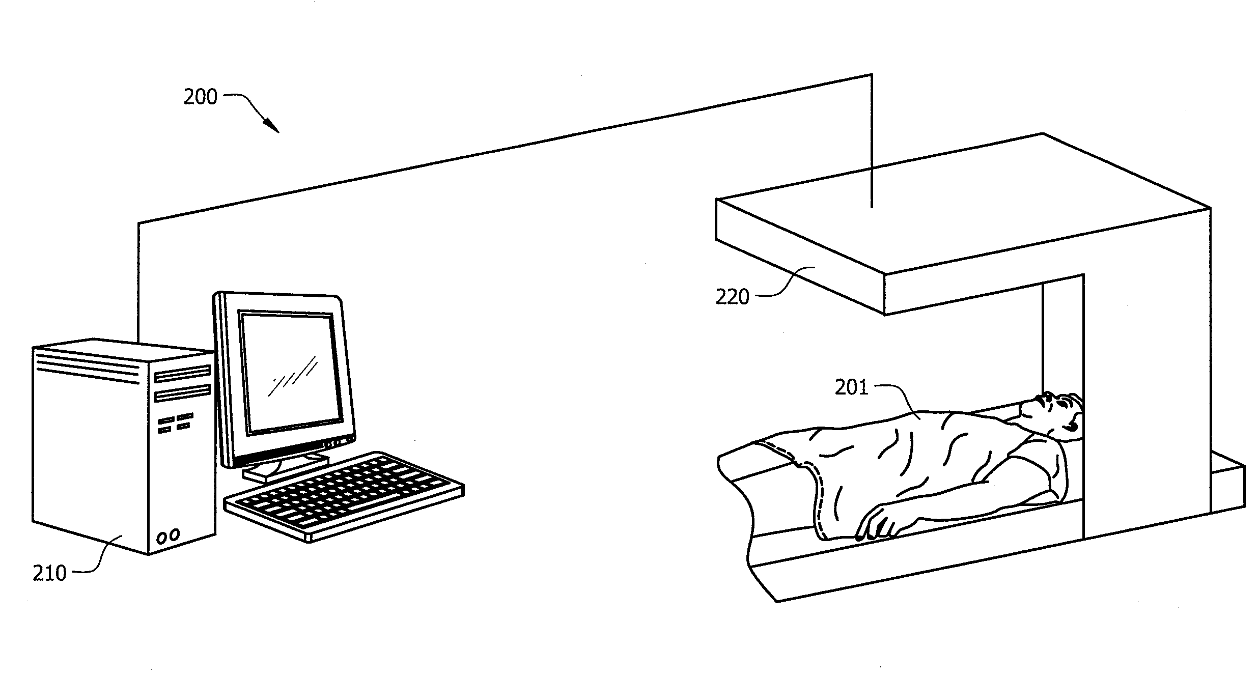
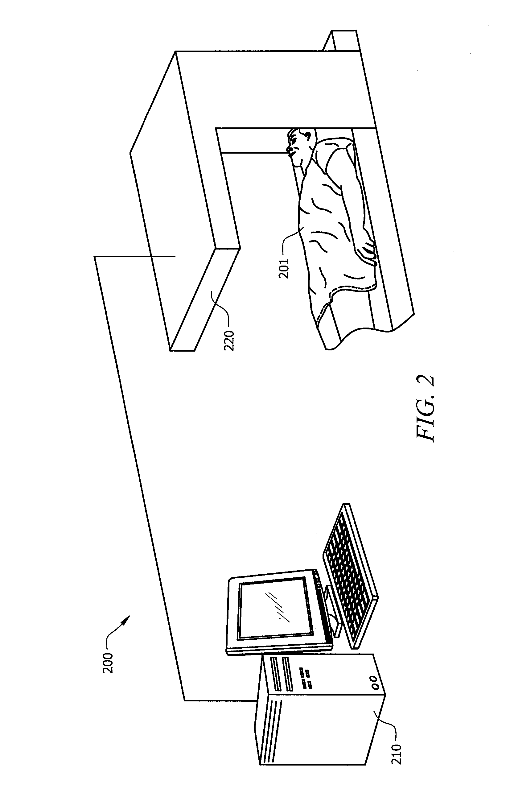
[0034]Directing attention to FIG. 2, an imaging system adapted according to an embodiment of the present invention is shown as imaging system 200. Imaging system 200 comprises processor based system 210 operatively coupled to transducer 220 for use in collecting imaging data with respect to subject 201. For example, subject 201 may comprise a patient for which imaging of a portion of the patient's body (e.g., hea...


PUM


Abstract

Systems and methods which implement an image reconstruction framework to incorporate complex motion of structure, such as complex deformation of an object or objects being imaged, in image reconstruction techniques are shown. For example, techniques are provided for tracking organ motion which uses raw time-stamped data provided by any of a number of imaging modalities and simultaneously reconstructs images and estimates deformations in anatomy. Operation according to embodiments reduces artifacts, increase the signal-to-noise ratio (SNR), and facilitates reduced image modality energy dosing to patients. The technology also facilitates the incorporation of physical properties of organ motion, such as the conservation of local tissue volume. This approach is accurate and robust against noise and irregular breathing for tracking organ motion.

Description

CROSS-REFERENCE TO RELATED APPLICATIONS[0001]This application claims priority to U.S. Provisional Patent Application No. 61 / 222,028 entitled “IMAGE RECONSTRUCTION INCORPORATING ORGAN MOTION,” filed Jun. 30, 2009, the disclosure of which is hereby incorporated herein by reference.TECHNICAL FIELD[0002]The disclosure relates generally to imaging and, more particularly, to reconstructing images which incorporate motion.BACKGROUND OF THE INVENTION[0003]Imaging techniques have been developed for studying organs where motion is present. For example, four-dimensional respiratory-correlated computed tomography (4D RCCT) has been widely used for studying organs where motion is associated with patient breathing. The current standard practice for reconstructing images with respect to such organ motion is to use phase binned images. However, the phase binning algorithm assumes that the patient has a periodic breathing pattern. When the patient's breathing is irregular as represented in the graph...

Claims


Application Information

Patent Timeline
Patent Type & Authority Applications(United States)
IPC IPC(8): G06K9/00
CPCA61B5/7289A61B6/00A61B8/00G06K9/00442G06T2211/412A61B6/5247G06F17/30247G06T11/006G06F16/583
Inventor JOSHI, SARANGHINKLE, JACOBSALTER, BILLFLETCHER, TOMWANG, BRIAN
Owner UNIV OF UTAH RES FOUND
Why Eureka
Try Eureka