
Combinational pixel-by-pixel and object-level classifying, segmenting, and agglomerating in performing quantitative image analysis that distinguishes between healthy non-cancerous and cancerous cell nuclei and delineates nuclear, cytoplasm, and stromal material objects from stained biological tissue materials

a quantitative image analysis and pixel-by-pixel technology, applied in image analysis, image enhancement, instruments, etc., can solve problems such as increasing the risk of breast cancer

Inactive Publication Date: 2013-07-16
TRIAD NAT SECURITY LLC
View PDF27 Cites 99 Cited by
  • Summary
  • Abstract
  • Description
  • Claims
  • Application Information

AI Technical Summary

Benefits of technology

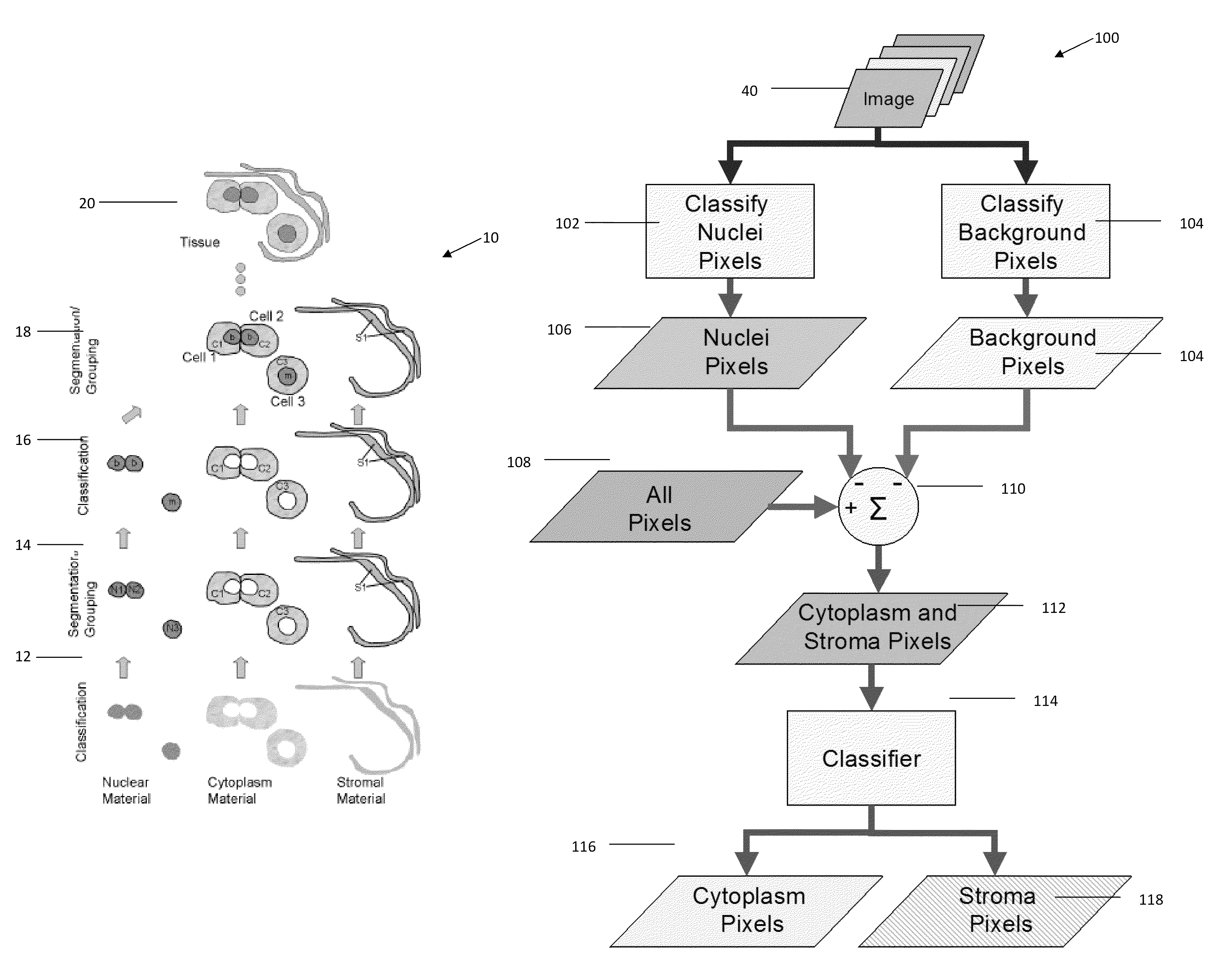
[0036]The present invention yields very good object- and image-level classification performances. More specifically, the object-level features as implemented in the present invention are versatile and general enough to elicit important information from even imperfectly segmented objects. Additionally, the use of non-nuclear features, namely features of cytoplasm and stroma, have good classification performance, often exceeding that of nuclei. Higher-level features display a potential to increase both object- and image-level classification performance.
[0038]The method further allows a user to mark up the image subsequent to the classification to re-classify said materials. Preferably, the markup is performed via a graphic user interface to edit designated regions in the image.
[0039]In a preferred embodiment, the image markup is used to develop one or more classifiers for specific classes of materials, wherein the classifiers are used to classify materials in subsequent imaging. In addition, the method may further include displaying the segmented regions, and allowing the user to markup the image to re-segment the identified classes.

Problems solved by technology

Breast cancer is now known to consist of a variety of diseases; complicating the diagnosis and treatment procedures are a large number of conditions that mimic breast cancer and may present an increased risk for developing breast cancer.

Method used


Image

  • Combinational pixel-by-pixel and object-level classifying, segmenting, and agglomerating in performing quantitative image analysis that distinguishes between healthy non-cancerous and cancerous cell nuclei and delineates nuclear, cytoplasm, and stromal material objects from stained biological tissue materials
  • Combinational pixel-by-pixel and object-level classifying, segmenting, and agglomerating in performing quantitative image analysis that distinguishes between healthy non-cancerous and cancerous cell nuclei and delineates nuclear, cytoplasm, and stromal material objects from stained biological tissue materials
  • Combinational pixel-by-pixel and object-level classifying, segmenting, and agglomerating in performing quantitative image analysis that distinguishes between healthy non-cancerous and cancerous cell nuclei and delineates nuclear, cytoplasm, and stromal material objects from stained biological tissue materials

Examples


Embodiment Construction

[0128]Referring more specifically to the drawings, for illustrative purposes the present invention is embodied in the apparatus generally shown in FIG. 1 through FIG. 70. It will be appreciated that the apparatus may vary as to configuration and as to details of the parts, and that the method may vary as to the specific steps and sequence, without departing from the basic concepts as disclosed herein.

1. OVERVIEW

[0129]1.1 Objectives

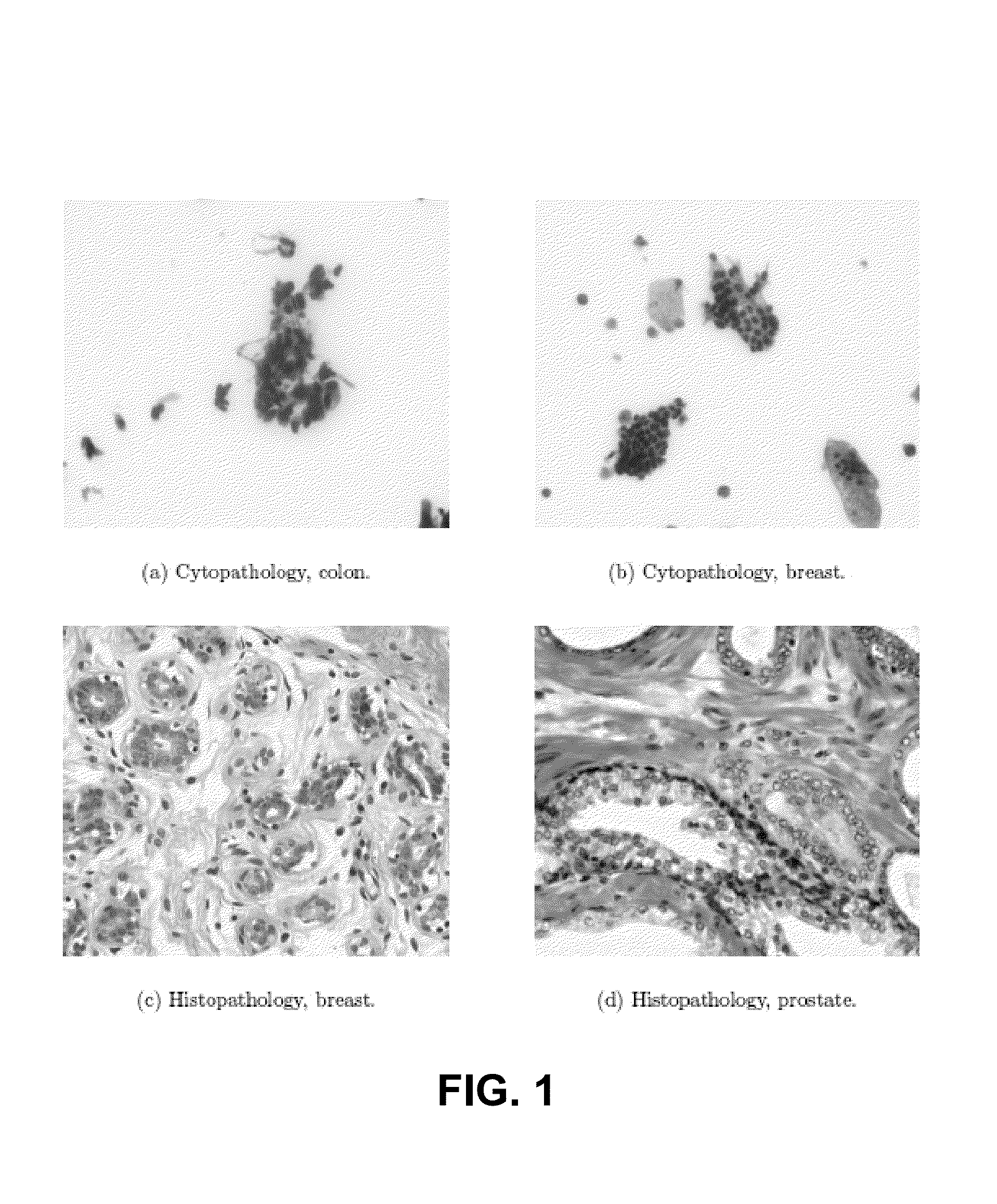
[0130]A primary objective of the present invention is the development of techniques for higher-level image analysis, i.e., object-level analysis. While the present invention may be employed in many applications, the discussion disclosed herein primarily is directed to systems and methods to assist in the quantification of breast cancer in histo / cytopathology imagery.

[0131]1.2 Pathology

[0132]1.2.1 Histo- and Cyto-Pathology

[0133]The following are definitions related to the disclosure herein described:

[0134]Pathology: the branch of medicine concerned with dis...


PUM


Abstract

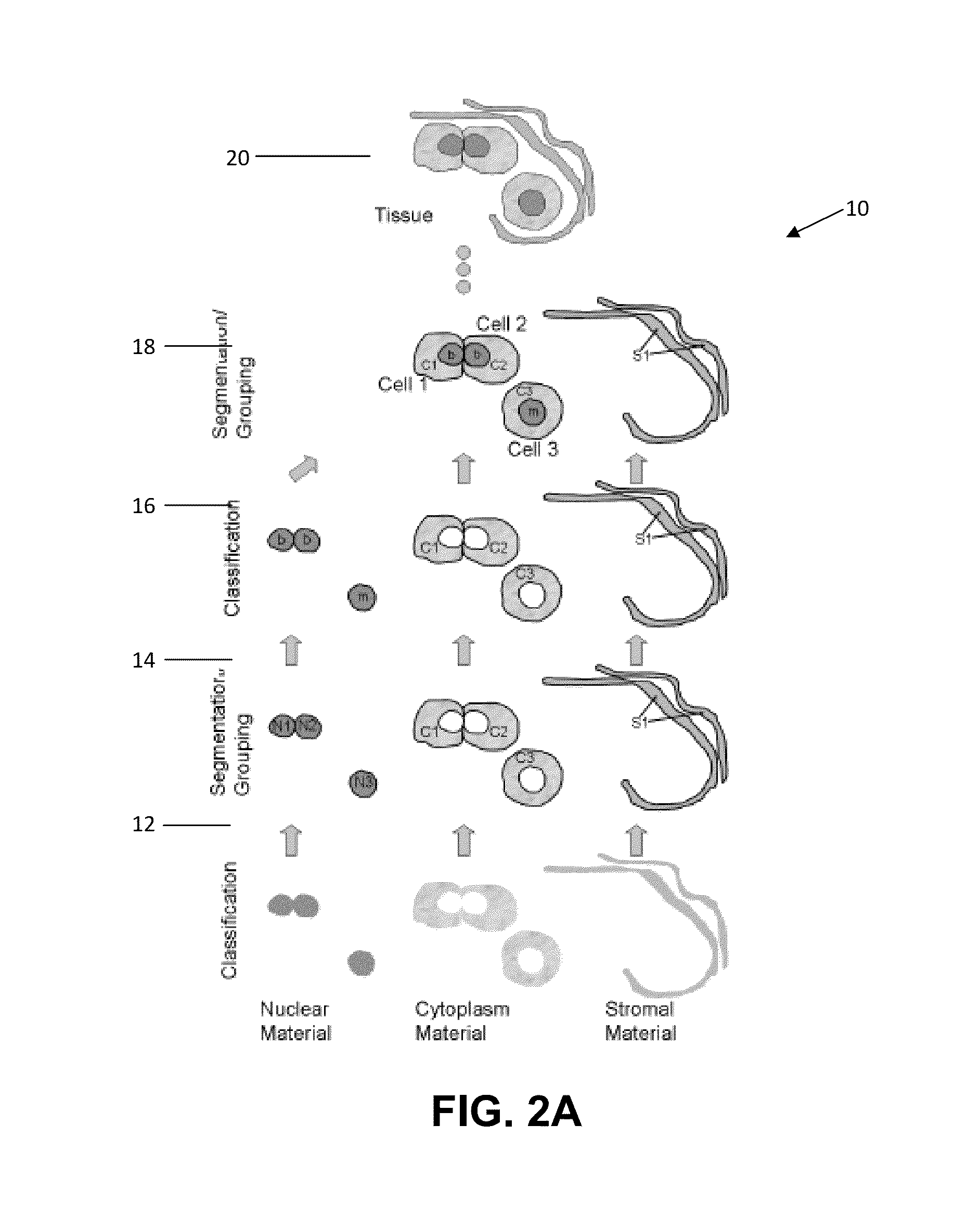
Quantitative object and spatial arrangement-level analysis of tissue are detailed using expert (pathologist) input to guide the classification process. A two-step method is disclosed for imaging tissue, by classifying one or more biological materials, e.g. nuclei, cytoplasm, and stroma, in the tissue into one or more identified classes on a pixel-by-pixel basis, and segmenting the identified classes to agglomerate one or more sets of identified pixels into segmented regions. Typically, the one or more biological materials comprises nuclear material, cytoplasm material, and stromal material. The method further allows a user to markup the image subsequent to the classification to re-classify said materials. The markup is performed via a graphic user interface to edit designated regions in the image.

Description

STATEMENT REGARDING FEDERALLY SPONSORED RESEARCH OR DEVELOPMENT[0001]This invention was made with Government support under Contract No. DE-AC52-06NA25396, awarded by the Department of Energy. The Government has certain rights in this invention.CROSS-REFERENCE TO RELATED APPLICATIONS[0002]Not ApplicableINCORPORATION-BY-REFERENCE OF MATERIAL SUBMITTED ON A COMPACT DISC[0003]Not ApplicableNOTICE OF MATERIAL SUBJECT TO COPYRIGHT PROTECTION[0004]A portion of the material in this patent document is subject to copyright protection under the copyright laws of the United States and of other countries. The owner of the copyright rights has no objection to the facsimile reproduction by anyone of the patent document or the patent disclosure, as it appears in the United States Patent and Trademark Office publicly available file or records, but otherwise reserves all copyright rights whatsoever. The copyright owner does not hereby waive any of its rights to have this patent document maintained in...

Claims


Application Information

Patent Timeline
Patent Type & Authority Patents(United States)
IPC IPC(8): G06K9/00G06V10/426G06V10/771G06V10/84
CPCG06K9/0014G06K9/469G06K9/6231G06K9/6296G06T7/0012G06T7/0081G06T2207/10056G06T2207/20141G06T2207/30024G06T2207/30068G06T7/11G06T7/187G06V20/695G06V10/426G06V10/771G06V10/84G06F18/2115G06F18/29
Inventor BOUCHERON, LAURA E.
Owner TRIAD NAT SECURITY LLC
Learn More