Spine labeling

a labeling and spine technology, applied in the field of spine labeling, can solve the problem that it is typically not possible for an untrained observer to accurately identify vertebrae, and achieve the effect of reducing the complexity of the algorithm, reducing the cost of accuracy and/or flexibility

Active Publication Date: 2013-08-13
PHILIPS MEDICAL SYST TECH
View PDF5 Cites 13 Cited by
  • Summary
  • Abstract
  • Description
  • Claims
  • Application Information

AI Technical Summary

Benefits of technology

[0017]Using one or more of the sieving procedures listed above, each aggregate is assigned a score that describes the likelihood of the aggregate to be a vertebra. At this stage, a significant portion of the aggregates in the image have been excluded from farther consideration. Optionally, exclusion of a significant portion of the aggregates from further consideration reduces a complexity and / or computational load in subsequent portions of the algorithm.
[0019]In an exemplary embodiment of the invention which employs MIS for aggregate segmentation, aggregates from different scales are merged, optionally hierarchically. In hierarchical merging a collection of aggregates containing a common point are evaluated to determine which of the aggregates is most likely to be a vertebra and / or be included in a chain being assembled. Optionally, only one of the aggregates is retained for consideration. In some embodiments of the invention, aggregate merging contributes to a reduction in complexity of the algorithm at the expense of accuracy and / or flexibility.

Problems solved by technology

In 3D medical images such as, for example, MRI and CT images, it is typically not possible for an untrained observer to accurately identify vertebrae.
This difficulty can be based in part on the similarity of a vertebra to its neighbors.

Method used


Image

  • Spine labeling
  • Spine labeling
  • Spine labeling

Examples


Embodiment Construction

Overview

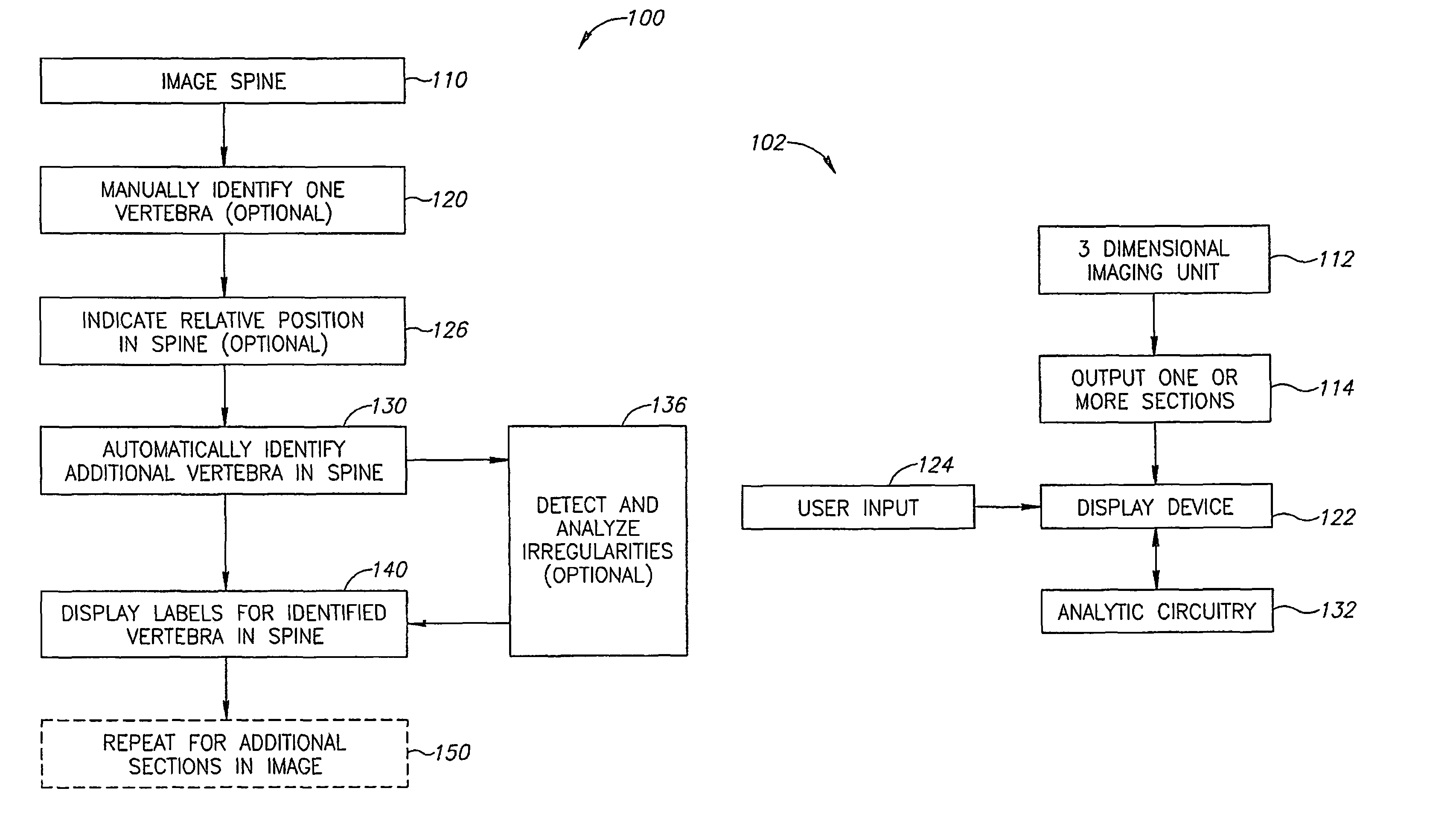
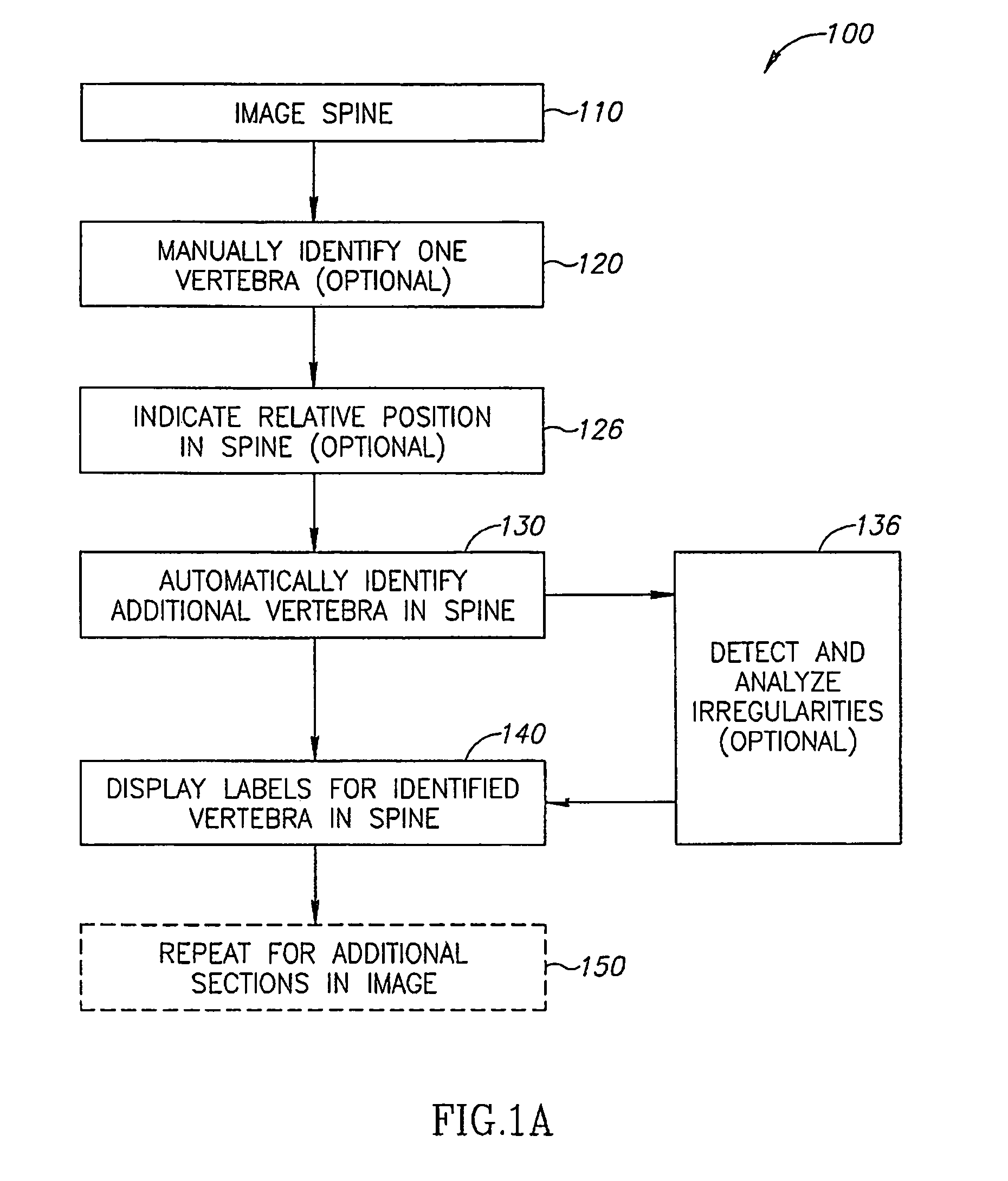
[0078]FIG. 1A is a simplified flow diagram illustrating an exemplary method 100 of labeling vertebrae in a medical image according to some embodiments of the invention.

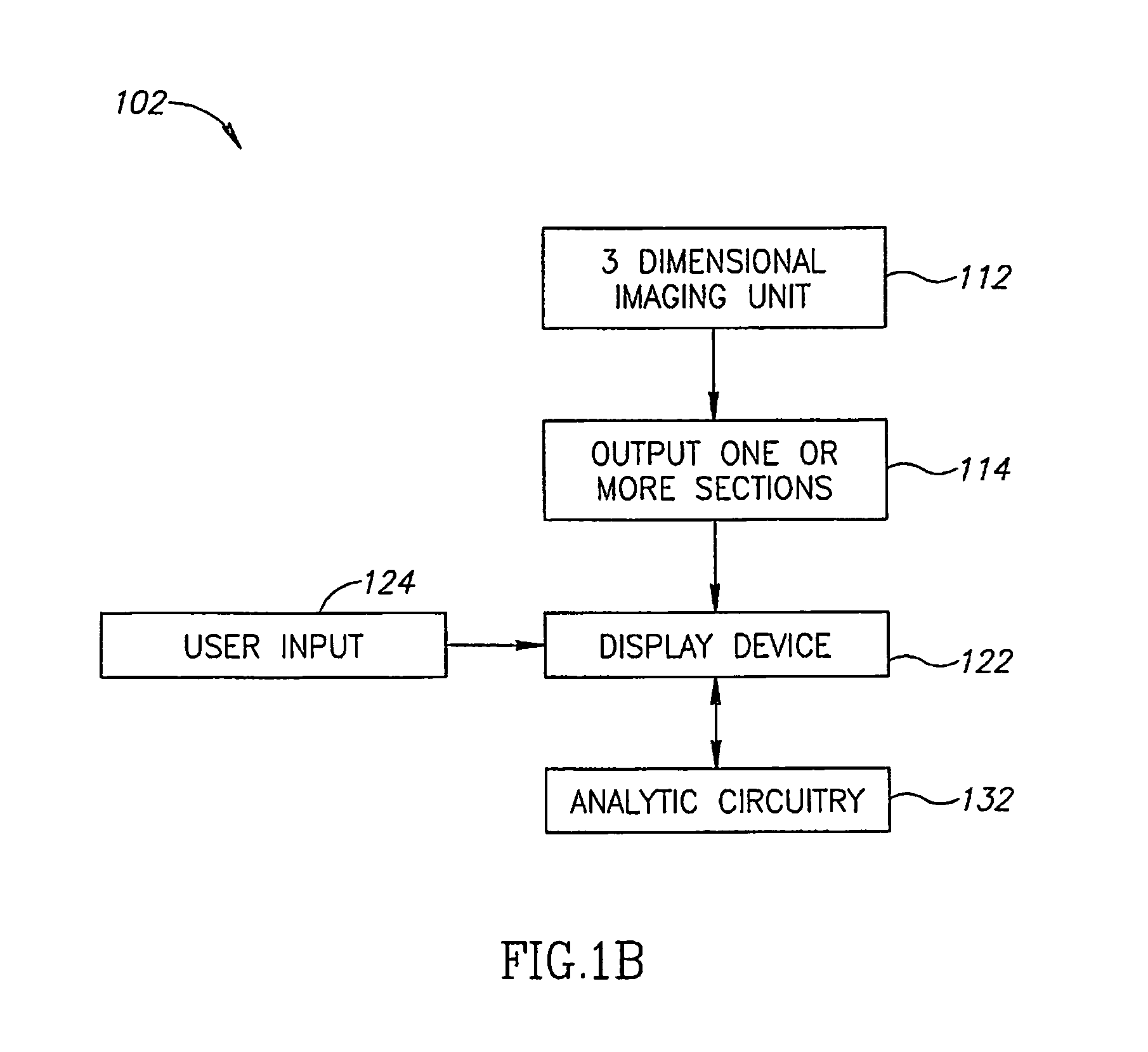
[0079]FIG. 1B is a schematic representation of an image analysis system 102 according to some exemplary embodiments of the invention.

[0080]Referring to FIGS. 1A and 1B, use of exemplary image analysis system 102 to perform an exemplary method 100 of labeling vertebrae is described. In general, the system and method attempt to assemble aggregates of pixels into a chain which represents a spine.

[0081]At 110 a spine is imaged using an imaging unit 112. In an exemplary embodiment of the invention, the imaging produces a 3D image. Optionally, imaging unit 112 is a magnetic resonance induction (MRI) unit or computerized tomography (CT) unit.

[0082]Imaging unit 112 provides one or more sections as output 114 which are optionally displayed on a display device 122 and / or presented as serial sections on a tangible media ...


PUM


Abstract

An image analysis system comprising: (a) an image input module adapted to receive a medical image; and (b) an analytic module adapted to segment the image, identify a plurality of vertebrae and label each vertebra.

Description

RELATED APPLICATION[0001]This application claims the benefit of priority from Israel Patent Application No. 179581 filed on Nov. 26, 2006, the contents of which are incorporated herein by reference.FIELD OF THE INVENTION[0002]The present invention relates to systems methods and / or user interfaces for identifying vertebrae in a medical image.BACKGROUND OF THE INVENTION[0003]In 3D medical images such as, for example, MRI and CT images, it is typically not possible for an untrained observer to accurately identify vertebrae. Even an experienced radiologist can have difficulty identifying specific vertebra within the spine according to the standard C1-C7, T1-T12, L1-L5, and S1-S5 naming convention. This difficulty can be based in part on the similarity of a vertebra to its neighbors. In some cases a trained radiologist can identify a certain exceptional vertebra, for example the C2 and / or the L5.SUMMARY OF THE INVENTION[0004]A broad aspect of the invention relates to a system which accep...

Claims


Application Information

Patent Timeline
Patent Type & Authority Patents(United States)
IPC IPC(8): G06K9/00
CPCG06T7/0081G06T2207/10081G06T2207/10088G06T2207/20016G06T2207/20072G06T2207/20101G06T2207/30012G06T7/11
Inventor PORAT, HADARAKERMAN, SHMUELPADAN, HADAS
Owner PHILIPS MEDICAL SYST TECH
Why Eureka
Try Eureka