Method and device for the segmentation of a lesion

a lesion and segmentation technology, applied in image data processing, character and pattern recognition, instruments, etc., can solve the problems of difficult to distinguish benign and malignant focal findings using their appearance in mammography images, and no reliable decision aid can be provided by the assistance system, so as to achieve reliable and robust effects

Inactive Publication Date: 2013-12-03
FRAUNHOFER GESELLSCHAFT ZUR FOERDERUNG DER ANGEWANDTEN FORSCHUNG EV
View PDF4 Cites 4 Cited by
  • Summary
  • Abstract
  • Description
  • Claims
  • Application Information

AI Technical Summary

Benefits of technology

[0008]The invention is therefore based on the object of providing a reliable and robust method for the segmentation of lesions.

Problems solved by technology

Distinguishing between benign and malignant focal findings using their appearance in a mammography image is difficult in practice as a mammography image is a two-dimensional transmitted-light recording of the three-dimensional breast, whereby optical superimpositions with breast tissue take place in the mammography image.
Optical superimpositions of benign focal findings with breast tissue are often difficult to distinguish from malignant focal findings in mammography images.
Known methods for segmentation in principle allow the separation of the lesion from the image background, but the separation is often too imprecise because of optical superimpositions, low contrast or high image noise, so no reliable decision aid can be provided by the assistance system.
Optical superimpositions, a low contrast or high image noise also impair the result of the segmentation in this method.

Method used


Image

  • Method and device for the segmentation of a lesion
  • Method and device for the segmentation of a lesion
  • Method and device for the segmentation of a lesion

Examples


Embodiment Construction

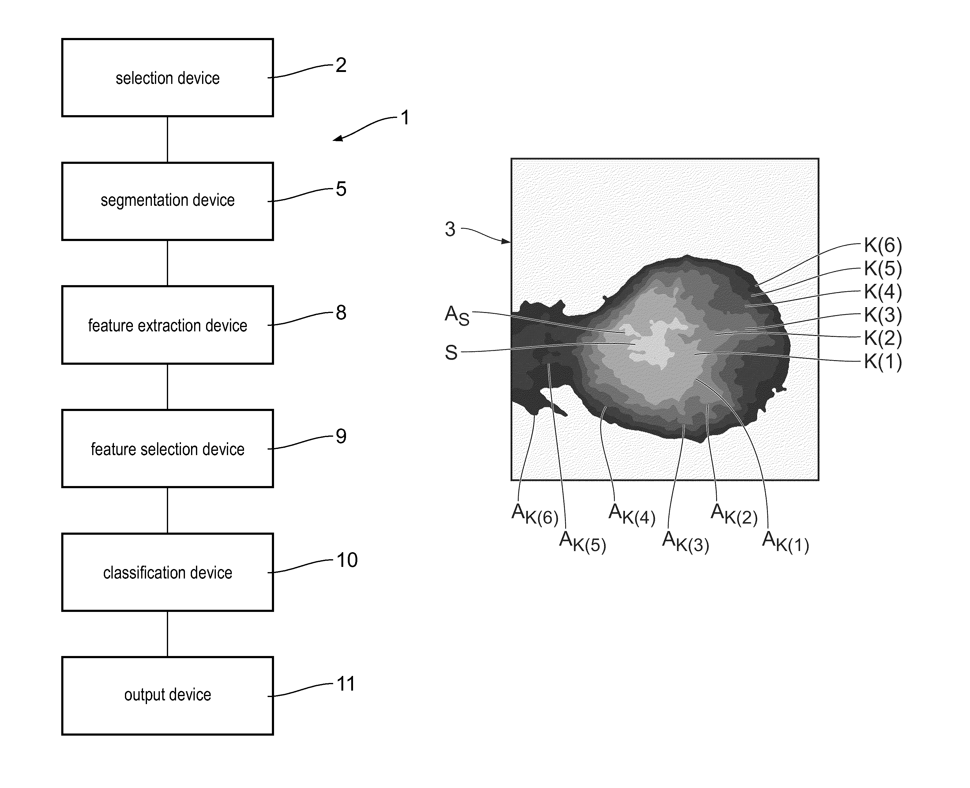
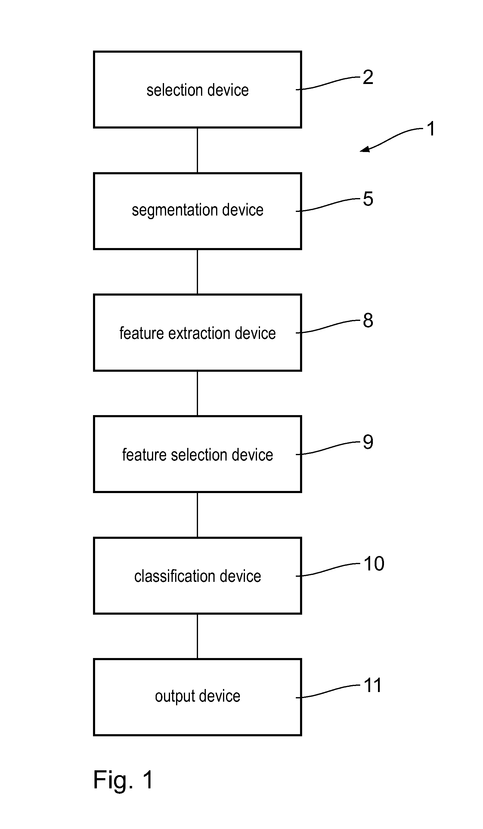
[0061]A first embodiment of the invention will be described below with reference to FIG. 1 to 10. An assistance system 1 is used to set up a decision aid to distinguish between a benign breast lesion and a malignant breast lesion.

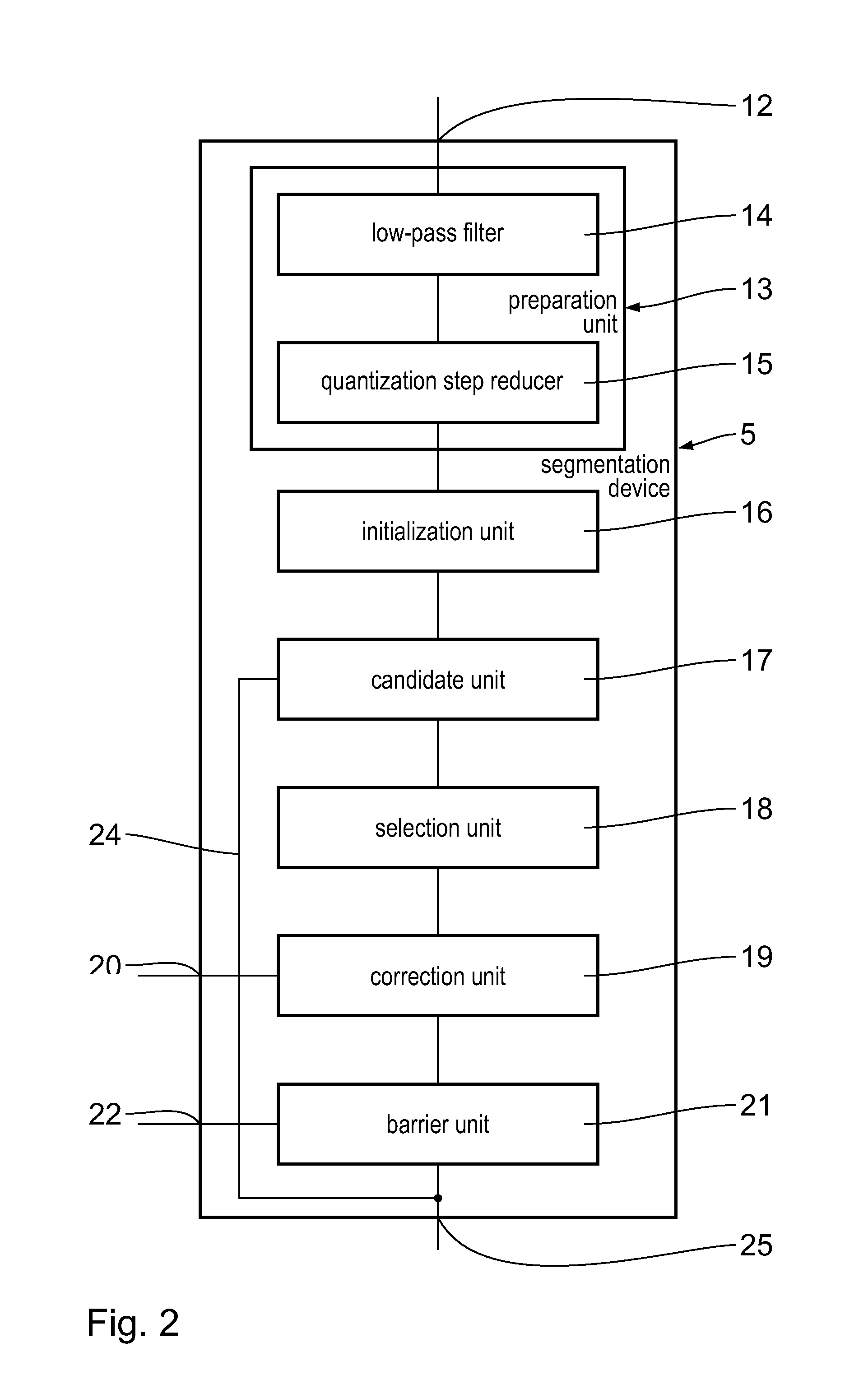
[0062]The assistance system 1 has a selection device 2. The selection device 2 is used to select an image region 3 of interest, which contains a lesion 4 in the form of a focal finding. The lesion will be designated below a focal finding 4. A device 5 for the segmentation of the focal finding 4 is arranged downstream of the selection device 2. The device will be designated a segmentation device 5 below. The segmentation device 5 is used to separate the focal finding 4 from an image background 6 of the image region 3. The segmentation device 5 supplies, as the result of the segmentation, a segmented focal finding 7, which ideally corresponds to the actual focal finding 4.

[0063]Arranged downstream of the segmentation device 5 is a feature extraction device 8....


PUM


Abstract

In a method and a device for the segmentation of a lesion, a quantity of candidate regions for the lesion is determined proceeding from a starting region by means of a candidate unit. The candidate regions are arranged substantially concentrically to the starting region. A candidate region closest to the lesion is selected automatically by means of a selection unit or manually by means of a correction unit from the quantity of candidate regions. The selected candidate region is a reliable and robust basis for a classification of the lesion.

Description

CROSS REFERENCE TO RELATED APPLICATIONS[0001]This application is a United States National Phase application of International Application PCT / EP2009 / 002198 and claims the benefit of priority under 35 U.S.C. §119 of German Patent Application DE 10 2008 016 807.6 filed Apr. 2, 2008, the entire contents of which are incorporated herein by reference.FIELD OF THE INVENTION[0002]The invention relates to a method and a device for the segmentation of a lesion.BACKGROUND OF THE INVENTION[0003]Breast cancer is the most frequent cancer disease in women in the western world. Early recognition of this disease improves the survival chances of the women affected. Digital X-ray mammography is the most effective technique for recognizing breast cancer tumors in early stages. An important group of breast lesions, which are to be found in digital mammography images are the so-called focal findings. Focal findings are local compressions, which are visible in at least two x-ray projections and are genera...

Claims


Application Information

Patent Timeline
Patent Type & Authority Patents(United States)
IPC IPC(8): G06K9/36
CPCG06T7/0012G06T7/0081G06T2207/10116G06T2207/30068G06T2207/30196G06T7/11
Inventor ELTER, MATTHIASBERGEN, TOBIAS
Owner FRAUNHOFER GESELLSCHAFT ZUR FOERDERUNG DER ANGEWANDTEN FORSCHUNG EV
Why Eureka
Try Eureka