Device for full thickness resectioning of an organ
a full-thickness resection and organ technology, applied in the direction of prosthesis, surgical staples, surgical forceps, etc., can solve the problems of lack of capability, size and structure, and deficiencies of known resection devices
- Summary
- Abstract
- Description
- Claims
- Application Information
AI Technical Summary
Benefits of technology
Problems solved by technology
Method used
Image
Examples
first embodiment
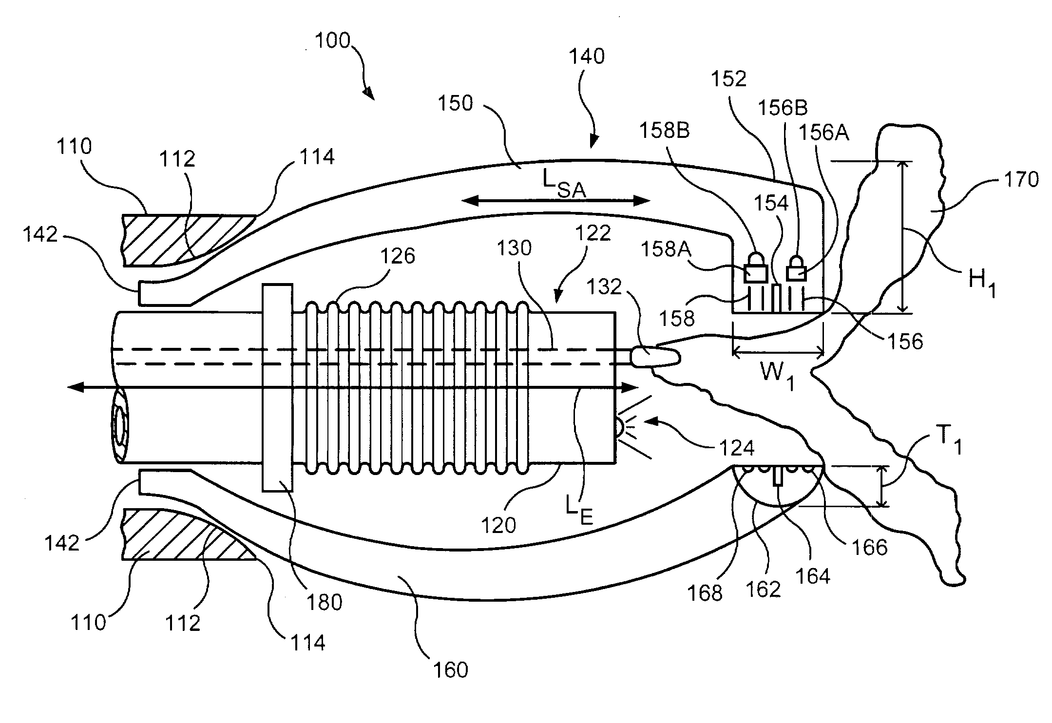
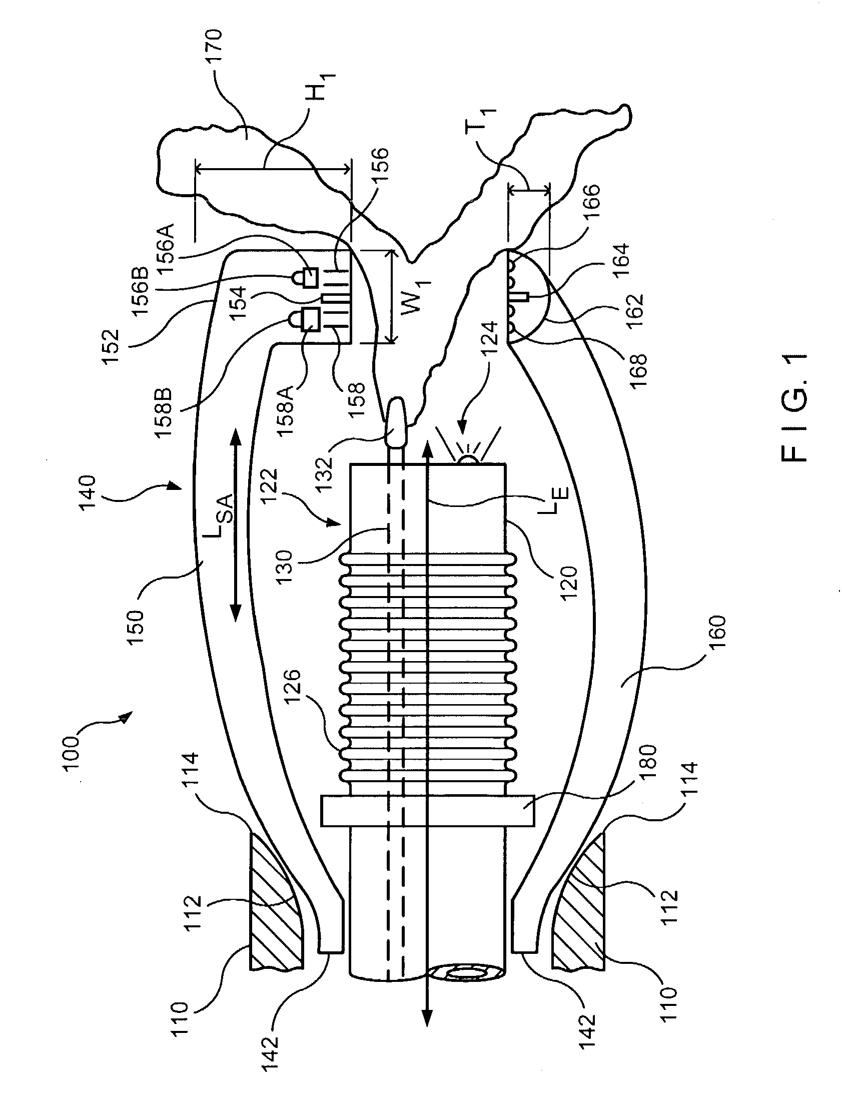
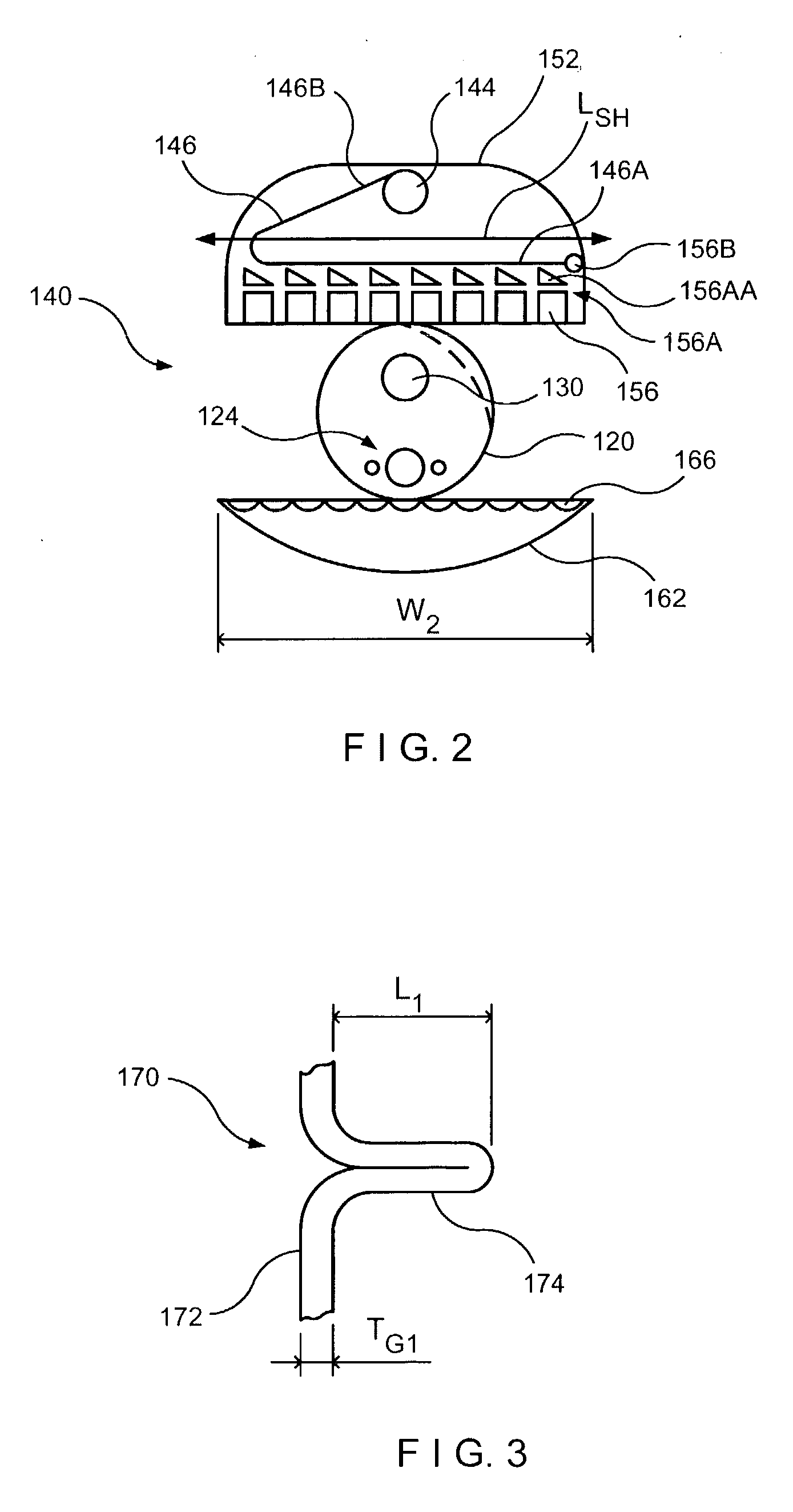
[0043]FIG. 1 illustrates a first embodiment for a full-thickness resection system 100 in accordance with the present invention. As can be seen in FIG. 1, full-thickness resection system 100 may include a closing cam 110, which may be associated with and / or actuated by a flexible shaft, a grasper 130, and a stapling mechanism 140. A flexible guide member 120, which in this embodiment is a flexible endoscope, is included within full thickness resection system 100 and grasper 130 may extend through a lumen included in the flexible endoscope 120. A stop 180 may be included on flexible endoscope 120. Closing cam 110 may be included on the distal ends of the flexible shaft or may be movable within the flexible shaft. Stapling mechanism 140 includes a stapling arm 150, which also includes a stapling head 152, and an anvil arm 160, which includes an anvil head 162. Each of these components of full-thickness resection system 100 will be described in further detail below. In utilizing full-th...
second embodiment
[0068]FIG. 12 illustrates an application where it is desirable that a grasper 230 grasps tissue 270 and pulls tissue 270 in a direction that is perpendicular to the longitudinal axis LE of the endoscope, which in this particular application, is more particularly defined as a deuodenoscope. Thus, in this embodiment for the endoscope, the deuodenoscope 220 may also include an optic 224 and a bending section 226. However, again, in this embodiment, the grasper 230 pulls the tissue 270 in a perpendicular direction to the longitudinal axis of the deuodenoscope, which is contrasted with the embodiment of FIG. 1 where the grasper 130 pulled the tissue in a direction that was parallel to the longitudinal axis of the endoscope 120. Thus, because the direction of pull of the tissue 270 is now perpendicular to the longitudinal axis of the deuodenoscope, it is not desirable that the stapling head be perpendicular to the longitudinal axis of the endoscope, as it was in the embodiment of FIG. 1, ...
third embodiment
[0072]FIGS. 14 and 15 illustrate a third embodiment for a full-thickness resection system 300 of the present invention that provides for perpendicular pulling of tissue within the stapling mechanism where the longitudinal axis of the stapling mechanism and the longitudinal axis of the endoscope are parallel.
[0073]As can be seen in FIG. 14, full-thickness resection system 300 includes a flexible shaft 310, a flexible endoscope 320, and a stapling mechanism 340. Flexible endoscope 320 has a longitudinal axis LE and stapling mechanism 340 has a longitudinal axis LSH which are parallel to each other. As discussed previously in the context of the other embodiments, stapling mechanism 340 includes a stapling head 352 and an anvil head (not visible in FIG. 14). Although not visible in FIG. 14, a grasper extends within a lumen included within flexible endoscope 320 and is movable on the longitudinal axis of flexible endoscope 320. Thus, the grasper extends from the distal end of the flexibl...
PUM
| Property | Measurement | Unit |
|---|---|---|
| Thickness | aaaaa | aaaaa |
| Pressure | aaaaa | aaaaa |
| Diameter | aaaaa | aaaaa |
Abstract
Description
Claims
Application Information
 - R&D
- Intellectual Property
- Life Sciences
- Materials
- Tech Scout
- Unparalleled Data Quality
- Higher Quality Content
- 60% Fewer Hallucinations
Browse by: Latest US Patents, China's latest patents, Technical Efficacy Thesaurus, Application Domain, Technology Topic, Popular Technical Reports.
© 2025 PatSnap. All rights reserved.Legal|Privacy policy|Modern Slavery Act Transparency Statement|Sitemap|About US| Contact US: help@patsnap.com


