Method for analyzing cervical lymph node metastasis, and tumor marker for head and neck cancer
a technology for cervical lymph nodes and tumor markers, applied in the field of cervical lymph node metastasis analysis, and tumor markers for head and neck cancer, can solve the problem of small detection limit of potential lymph node metastases
- Summary
- Abstract
- Description
- Claims
- Application Information
AI Technical Summary
Benefits of technology
Problems solved by technology
Method used
Image
Examples
example 1
[0049][Samples]
[0050]Samples in this Example were metastasized lymph nodes (7 samples) derived from patients of head and neck squamous cell carcinoma. For control samples, a lymph node (1 sample) and salivary glands (5 samples) derived from patients bearing no cancer were used. It should be noted that for use of the specimens in the present study, each patient and his / her family were fully informed, and thus a written consent was obtained.
[0051][Assay]
[0052]After homogenizing mechanically the respective samples, total RNA was extracted and purified by using Isogen (Nippon Gene, Toyama, Japan). 1 μg of the total RNA was amplified by using Cheluminescent RT-IVT Labeling Kit (Applied Biosystems, FosterCity, Calif.), thereby synthesizing digoxigenin (Roche Diagnostics, Basel, Switzerland) labeled cRNA. After hybrizing the synthesized cRNA with human genome survey microarray (Applied Biosystems), it was washed by using Chemiluminescent Detection Kit (Applied Biosystems) and, after color ...
example 2
[0058][Samples]
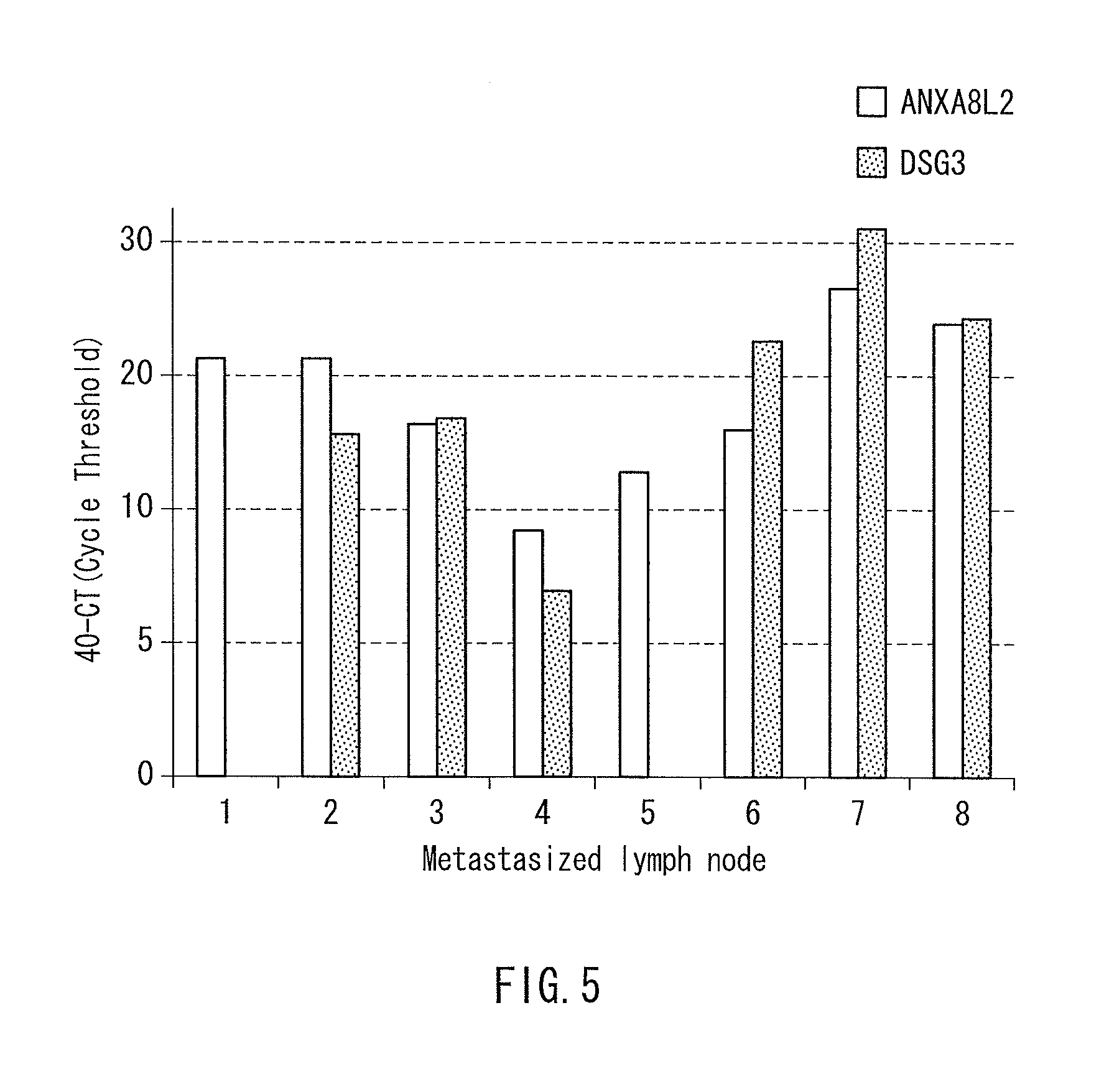
[0059]Samples in this Example were 8 specimens of lymph nodes. Regarding the specimens, the metastasis had been determined as negative in a conventional metastasis genetic test for detecting Cytokeratin19 (CK19) mRNA, but metastasis of head and neck cancer had been clarified by a microscopic examination on pathological tissues. It should be noted that for use of the specimens in the present study, each patient and his / her family were fully informed, and thus a written consent was obtained.
[0060][Use of Tumor Marker]
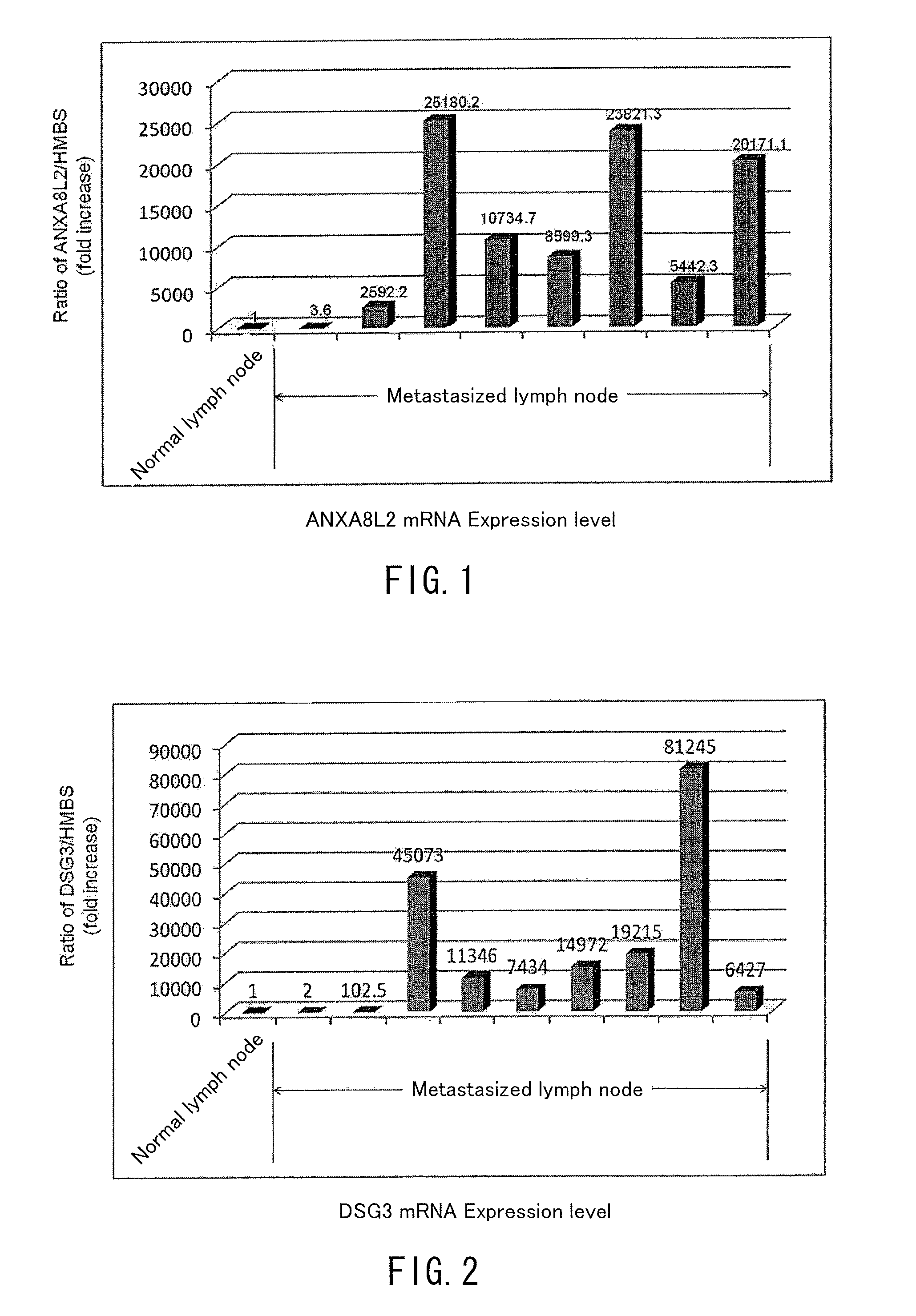
[0061]mRNA expression levels of the genes of SEQ ID NOS: 6 and 9 in the sequence listing (respectively ANXA8L2 and DSG3(PVA) genes) were measured for the samples, and the expression levels were compared with the expression level at a normal lymph node (lymph node derived from a patient bearing no cancer). The measurement of expression level was conducted by the realtime quantitative RT-PCR method similarly to Example 1.
[0062]The results are shown in FIG. 5....
example 3
[0063][Samples]
[0064]The samples in this Example were newly-prepared metastasized lymph nodes (12 samples) derived from head and neck squamous cell carcinoma patients different from those for Examples 1 and 2, and control samples were newly-prepared lymph nodes (7 samples) derived from patients bearing no cancer. It should be noted that for use of the specimens in the present study, each patient and his / her family were fully informed, and thus a written consent was obtained.
[0065][Use Of Tumor Marker]
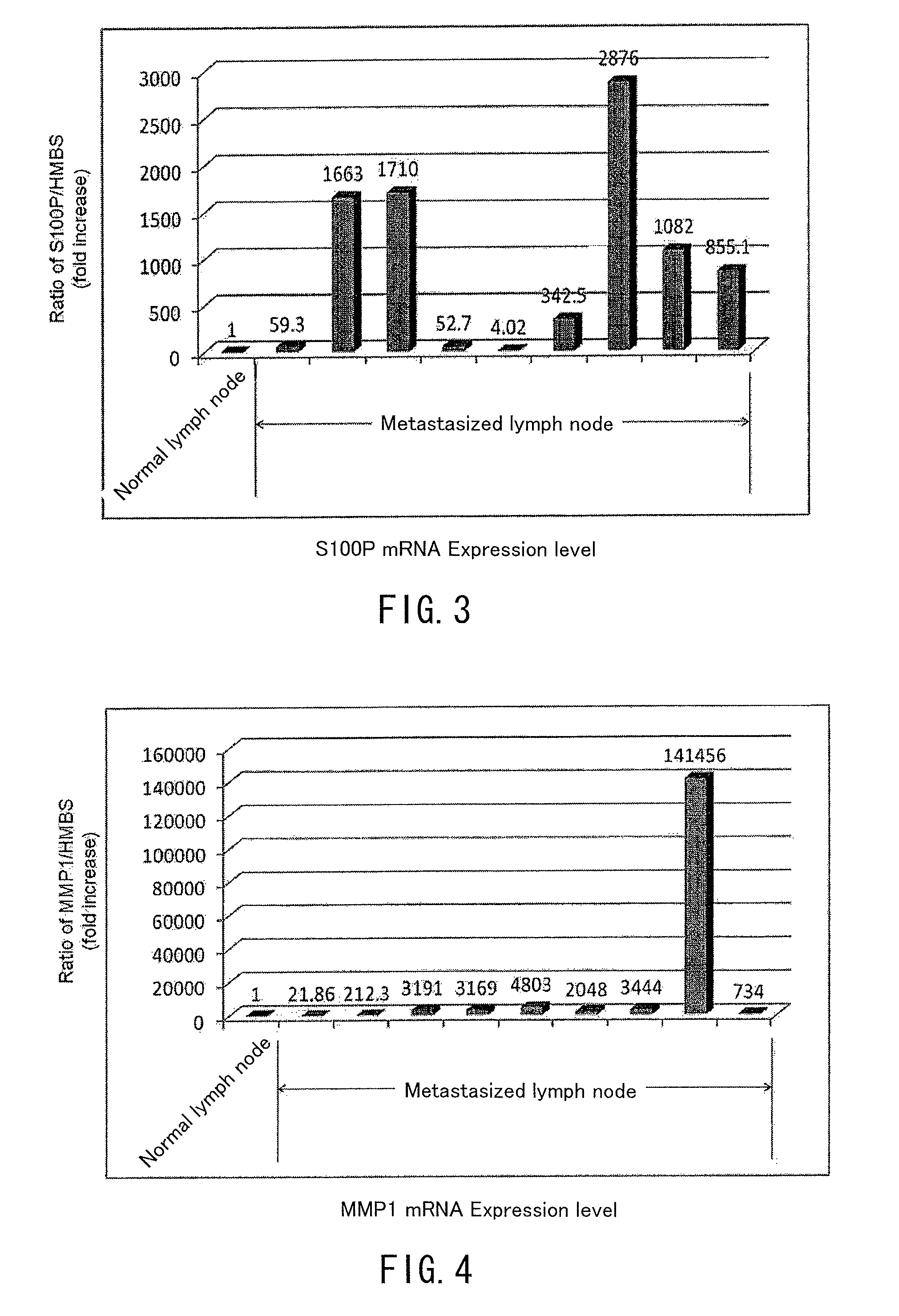
[0066]mRNA expression levels of the genes of SEQ ID NOS: 6, 9, 4, 2, 23, 13 and 32 in the sequence listing (respectively ANXA8L, DSG3(PVA), KRT-1, KRT-6A, MMP1, S100P and ARSI genes) were measured for the samples, and the expression levels were compared with the expression level at control samples. The measurement of expression level was conducted by the realtime quantitative RT-PCR method similarly to Example 1.
[0067]The results are shown in FIGS. 6 and 7. As illustrated in FIG. 6, for...
PUM
| Property | Measurement | Unit |
|---|---|---|
| diameter | aaaaa | aaaaa |
| real time quantitative RT- | aaaaa | aaaaa |
| size | aaaaa | aaaaa |
Abstract
Description
Claims
Application Information
 - R&D
- Intellectual Property
- Life Sciences
- Materials
- Tech Scout
- Unparalleled Data Quality
- Higher Quality Content
- 60% Fewer Hallucinations
Browse by: Latest US Patents, China's latest patents, Technical Efficacy Thesaurus, Application Domain, Technology Topic, Popular Technical Reports.
© 2025 PatSnap. All rights reserved.Legal|Privacy policy|Modern Slavery Act Transparency Statement|Sitemap|About US| Contact US: help@patsnap.com


