Kits for and methods of differential staining of cervical cancer cells and/or tissues

a technology of cervical cancer cells and kits, applied in the field of kits and methods of differential staining of cervical cancer cells and/or tissues, can solve the problems of becoming a major health hazard for the patient with cervical cancer

Active Publication Date: 2015-11-10
ZETIQ TECH
View PDF43 Cites 0 Cited by
  • Summary
  • Abstract
  • Description
  • Claims
  • Application Information

AI Technical Summary

Problems solved by technology

Epidemiological research conducted in 2003 in the central and western regions of China showed that cervical cancer is becoming a major health hazard for women in the countryside, with nearly 100,000 cases of cervical cancer being diagnosed annually, with approximately 20,000 deaths in 2001.
However, the Papanicolaou staining procedure, as well as all other cytological staining procedures, does not reveal any tinctorial selectivity for malignant cells, and the assessment of pathologically relevant traits is performed by means of interpretation of cellular morphology, which usually requires highly qualified and experienced cytopathologists.

Method used


Image

  • Kits for and methods of differential staining of cervical cancer cells and/or tissues
  • Kits for and methods of differential staining of cervical cancer cells and/or tissues
  • Kits for and methods of differential staining of cervical cancer cells and/or tissues

Examples


example 1

Diagnosing Cervical Cancer by Cervical Histology

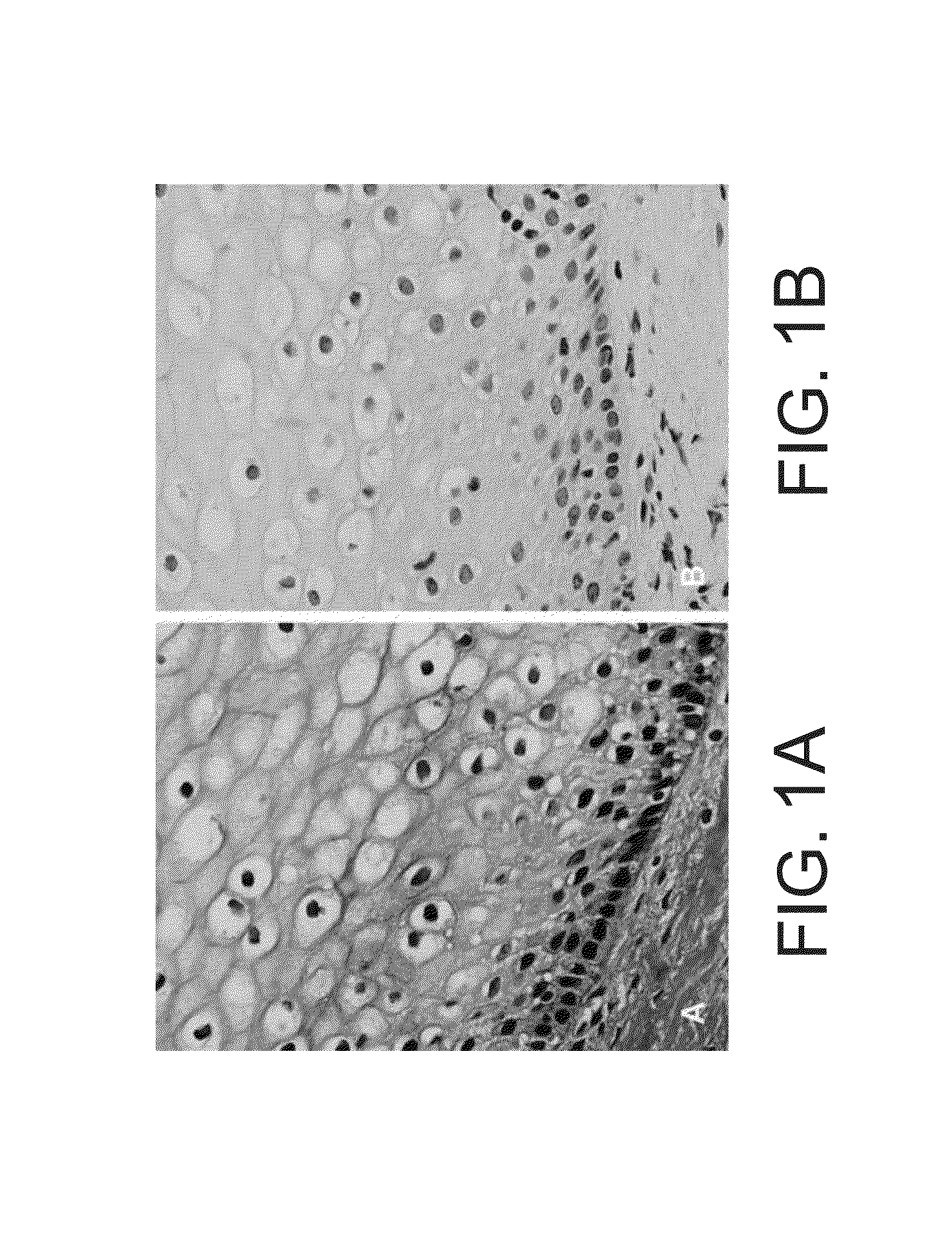
[0384]To establish the feasibility of the staining method according to some embodiments of the invention in detecting cervical cancer in cervical biopsies, optimize staining procedure, establish a correlation of atypical cell morphology with the presence of red color in those atypical cells, optimize and validate the ability of the staining method of some embodiments of the invention to detect cervical cancer and determine the sensitivity of the staining method of some embodiments of the invention to detect cervical cancer, the present inventors have conducted a single center open labeled study. The study was conducted at the Meir Medical Center, Kfar Saba, Israel, and included up to 60 cervical biopsies.

[0385]Study Outline

[0386]Biopsy samples with known diagnosis were received from Meir medical center pathology department, and transferred to a pathology laboratory to prepare multiple slides from each biopsy. The study is an open label...

example 2

Diagnosis of Cervical Cancer by Staining Conventional Cervical Smears

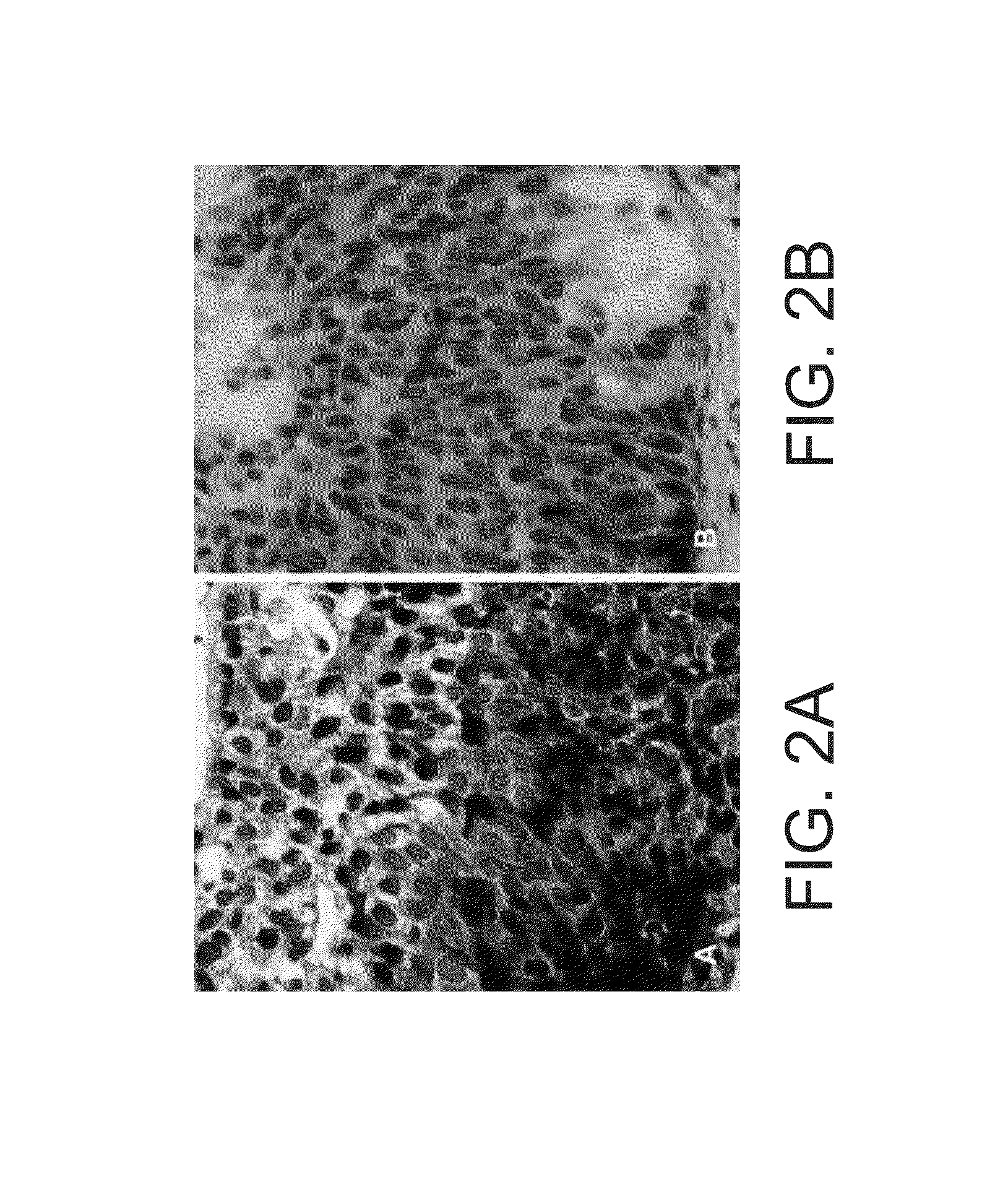
[0484]The study was designed to optimize and validate the ability of staining method of the invention to detect cervical cancer using a cervical smear. The study is an open label learning and calibration study of cervical smears with known pathology, intended for the optimization of the staining procedure and testing its feasibility in detecting cervical cancer.

[0485]Materials and Experimental Methods

[0486]Cervical smears on microscopical slides.

[0487]Protocol of Staining Conventional Cervical Smears:

[0488]Slides were washed in 100% Ethanol (EtOH) for 20 minutes, followed by a wash in double distilled water (DDW). Slides were then stained with Hematoxylin (ready for use solution) for 1 minute, rinsed under running Tap water for 2 minutes and washed in DDW for 10 seconds. Slides were then incubated with the Ficus elastics plant extract for 4 minutes, washed in DDW and incubated with 0.5% NF (New Fuchsin) in 20% EtOH...

example 3

Diagnosis of Cervical Cancer by Staining Liquid Based Slides (Thin-Prep™ Slides) and Comparison to Other Diagnostic Methods

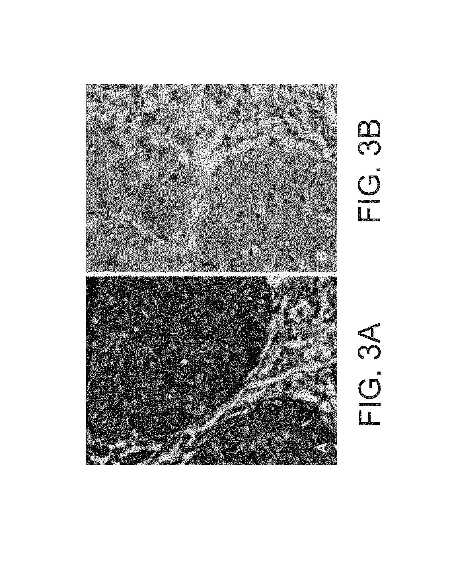
[0498]This study was designed to validate the staining method and kits of some embodiments of the invention as an adjunct to the diagnosis of cervical neoplastic processes in cervical cytology. This open labeled study was conducted on cervical samples with a known diagnosis, collected at Meir medical center or at Maccabi HMO in Israel. To obtain cervical samples with known diagnosis, subjects were recruited to the study upon visiting the medical facility (e.g., the colposcopy clinic) for follow up examinations following a suspected atypia. This protocol further served to increase the percent of subjects with dysplasia, which are of low prevalence in the general population. Accordingly, the percentage of subjects with dysplasia in this study was high.

[0499]The first part of this study entailed calibration and optimization of the staining process towards cervical ...


PUM

PropertyMeasurementUnit
thicknessaaaaaaaaaa
thicknessaaaaaaaaaa
thicknessaaaaaaaaaa

Abstract

Provided are methods and kits for staining cervical cell sample by contacting the cervical cell sample with a Ficus elastics plant extract, staining the cervical cell sample with New Fuchsin, and staining the cervical cell sample with Light Green or Fast Green. Also provided are method of diagnosing a pre-malignant or a malignant cervical tumor in a subject, by staining the cervical cell sample and identifying at least one cervical cell having a red cytoplasm above a pre-determined threshold, wherein presence of the at least one cervical cell having the red cytoplasm above the pre-determined threshold is indicative of a non- or less-differentiated cell as compared to a normal cervical cell, thereby diagnosing the pre-malignant or a malignant cervical tumor in the subject.

Description

REFERENCE TO CO-PENDING APPLICATIONS[0001]Priority is claimed as a U.S. national entry under 35 U.S.C. 371 of PCT / IL2010 / 000399, filed on May 17, 2010; which claims priority to U.S. provisional patent application Ser. No. 61 / 213,228, filed on May 19, 2009.FIELD AND BACKGROUND OF THE INVENTION[0002]The present invention, in some embodiments thereof, relates to kits and methods of differential staining of cervical cancer cells and / or tissues.[0003]Cervical cancer is the second most common cancer in women worldwide and is a leading cause of cancer-related death in women in underdeveloped countries. Worldwide, it is estimated that approximately 473,000 cases of cervical cancer are diagnosed each year, with about 253,500 deaths per year.[0004]Routine cervical cancer screening of the population by cervical vaginal smears has significantly decreased the incidence of cervical cancer in Western countries. In the United States, in 1998, about 12,800 women were diagnosed with cervical cancer a...

Claims


Application Information

Patent Timeline
Patent Type & Authority Patents(United States)
IPC IPC(8): G01N33/574
CPCG01N33/57411
Inventor IDELEVICH, PAVELELKELES, ADITERKIELTAUB, DOVEYAL, AMI
Owner ZETIQ TECH
Why Eureka
Try Eureka