
Intraocular lens

a technology of intraocular lens and lens sclera, which is applied in the field of intraocular lens, can solve the problems that the adaptability (or specialization) of intraocular lens for fixation into the sclera is not sufficiently developed

Active Publication Date: 2016-09-20
CHUKYO MEDICAL
View PDF9 Cites 2 Cited by
  • Summary
  • Abstract
  • Description
  • Claims
  • Application Information

AI Technical Summary

Benefits of technology

[0006]In this method, for example, there is no need to learn an intraocular lens suturing technique necessary for the method of suturing the intraocular lens. Further, it is reported that, when this method is used, the intraocular lens may be disposed at the center position of the eye with high precision so as to improve the lens fixing stability (particularly, the axial stability of the lens) and hence an effect is obtained in which the lens is not easily inclined.
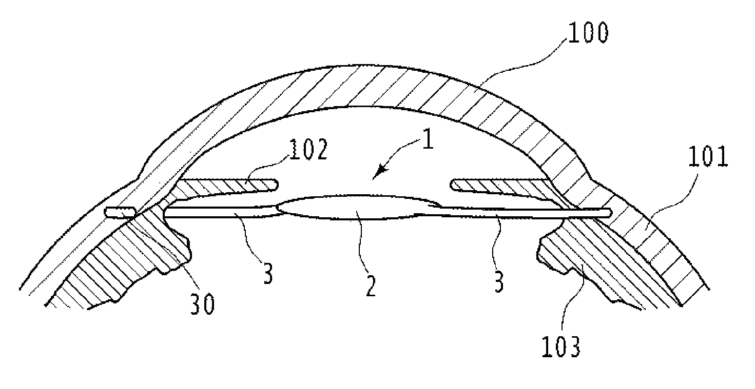
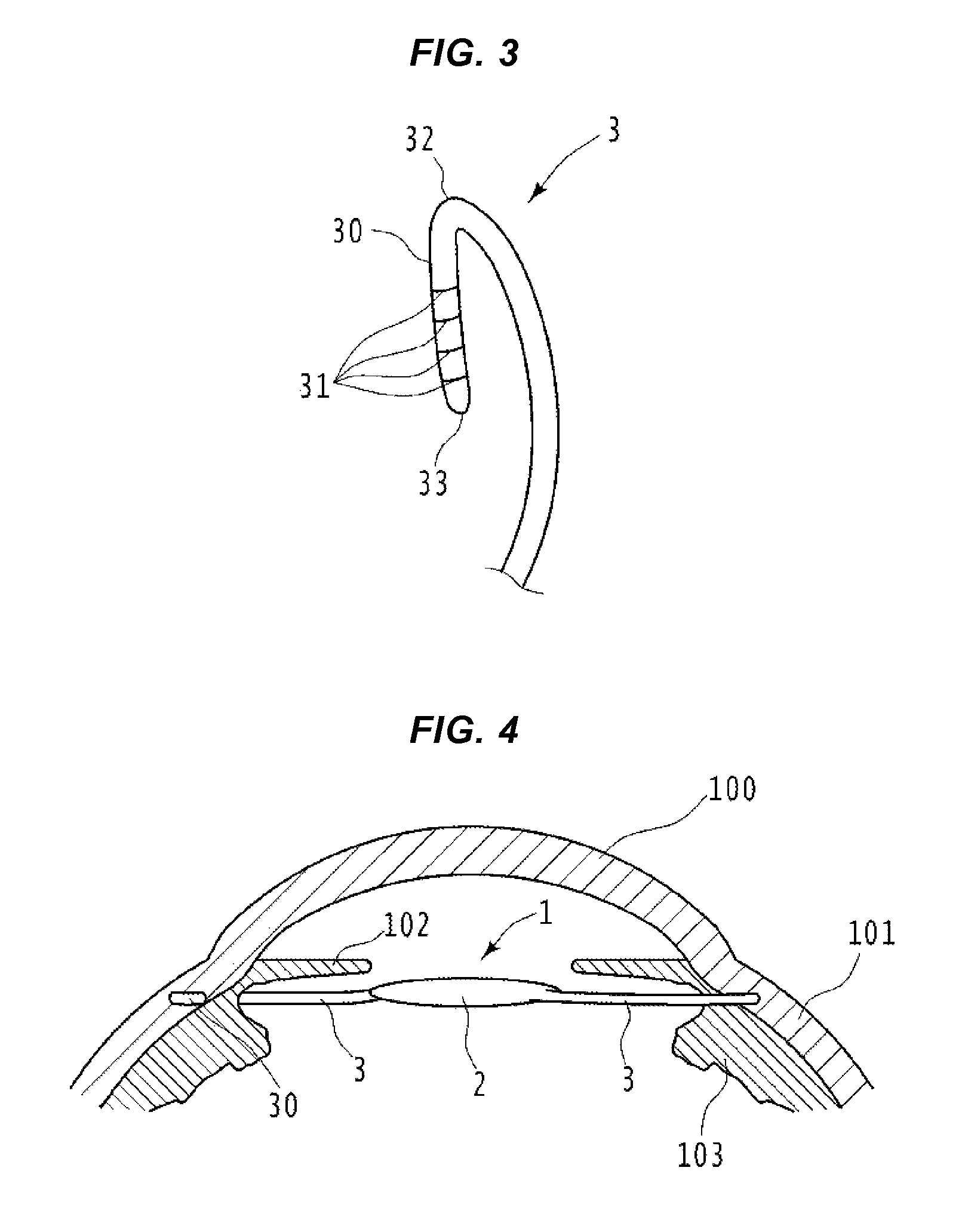
[0011]The folding-back portion may be folded back at an angle at which the folding-back portion is insertable into a tunnel-shaped hole portion formed inside the sclera along with a part of the support portion other than the folding-back portion. Accordingly, since the folding-back portion is insertable into the tunnel-shaped hole portion formed inside the sclera, the support portion is strongly restrained inside the sclera in a manner such that the folding-back portion inserted into the sclera is hooked to a certain portion inside the sclera. Thus, the intraocular lens is reliably fixed.
[0012]Further, when the folding-back portion is inserted into a tunnel-shaped hole portion formed inside the sclera along with a part of the support portion other than the folding-back portion, the length of the folding-back portion may be adjusted to the length in which the entire folding-back portion is accommodated inside the hole portion. Accordingly, since the entire folding-back portion may be accommodated in the tunnel-shaped hole portion formed inside the sclera, the support portion is strongly restrained inside the sclera in a manner such that the folding-back portion inserted into the sclera is hooked to a certain portion inside the sclera. Thus, the intraocular lens is reliably fixed.
[0014]Further, the support portion may be formed in a plurality of loop shapes extending from the limbus of the lens portion so as to be symmetrical to each other with respect to the visual axis, and the folding-back portion may be formed in each of the front ends of all support portions. Accordingly, since the plurality of loop-shaped support portions are strongly restrained inside the sclera by the folding-back portions of the front ends, the intraocular lens may be reliably fixed.
[0015]Further, the support portions may be formed in two loop shapes extending from the limbus of the lens portion so as to be symmetrical to each other with respect to the visual axis and the folding-back portion may be formed in the front ends of two support portions. Accordingly, since two loop-shaped support portions are strongly restrained inside the sclera by the folding-back portions of the front ends, the intraocular lens may be reliably fixed.

Problems solved by technology

In contrast, the intraocular lens appropriate (or specialized) for the fixation into the sclera is not sufficiently developed.
This challenge is not limited to the method of fixing the intraocular lens into the sclera, and also corresponds to the challenge for all treatments of fixing the intraocular lens to a part of the sclera like the outer surface of the sclera.

Method used


Image

  • Intraocular lens
  • Intraocular lens
  • Intraocular lens

Examples


Embodiment Construction

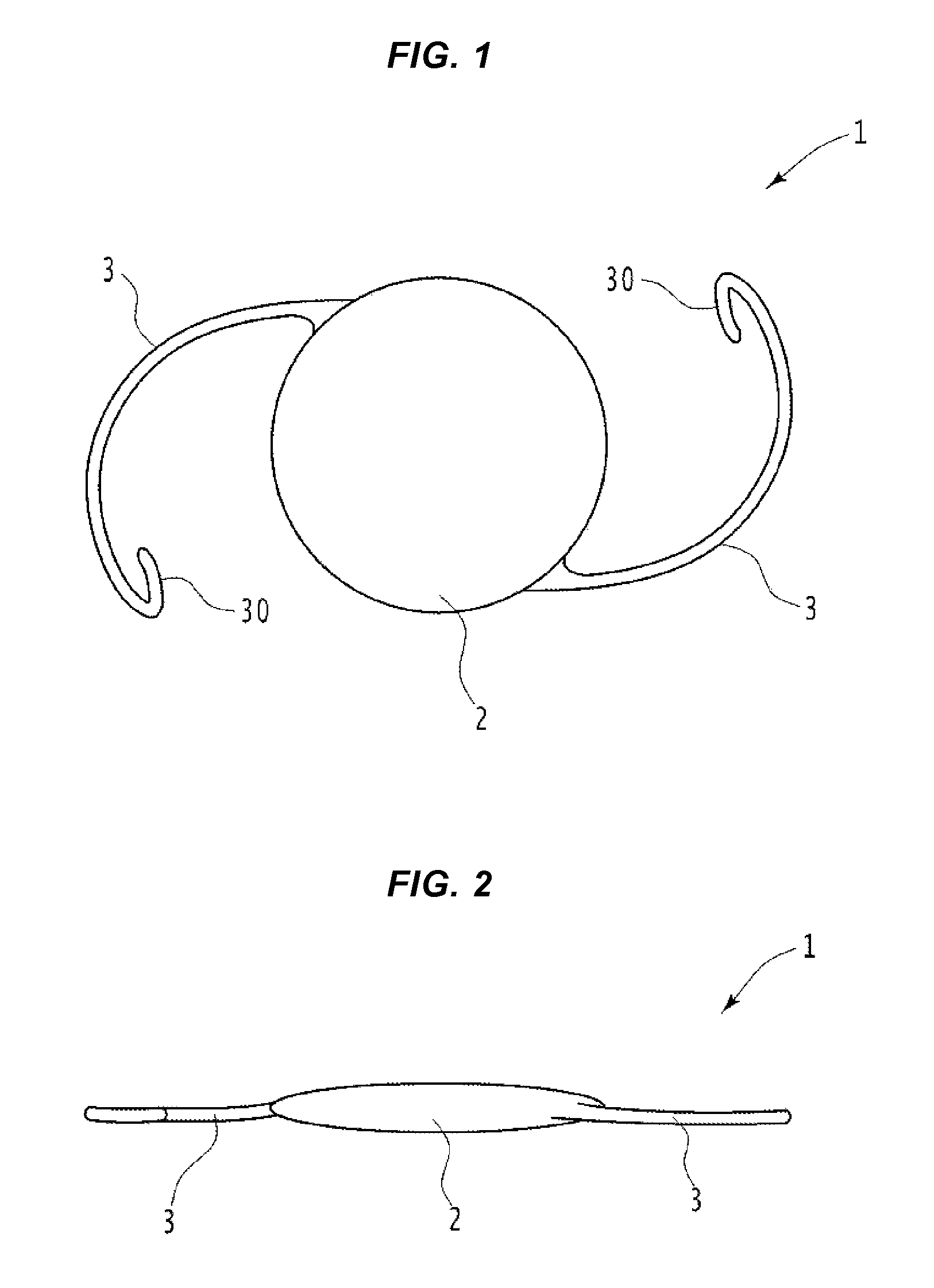
[0024]Hereinafter, an embodiment of the invention will be described by referring to the drawings. First, FIGS. 1 to 3 illustrate an intraocular lens 1 of an embodiment of the invention. FIG. 1 is a front view of the intraocular lens 1, and FIG. 2 is a side view thereof (the description on the direction of the side surface, the front surface, or the like indicates the direction (the side surface, the front surface, or the like) in a face (or an eye) of a patient having an eye into which the intraocular lens is fixed).

[0025]The intraocular lens 1 mainly includes a lens 2 and a support portion 3. For example, the lens 2 is disposed at the rear section (or an eye rear section, that is, an area behind the iris) after an eye lens which becomes cloudy white due to the cataract is extracted (entirely or partially extracted) from the patient's eye so that the lens carries out the lens function of the extracted eye lens.

[0026]The support portion 3 is a portion that supports the lens 2 to the ...


PUM


Abstract

Provided is an intraocular lens including a lens and a pair of right and left loop-shaped support portions, and a folding-back portion is provided in the front end of each support portion. A sclera tunnel is formed in the circumferential direction at a position of a depth corresponding to the half of the thickness of the sclera in two symmetrical positions with respect to the visual axis in a portion adjacent to a limbus of the sclera. The front end of the support portion is extracted from the ciliary sulcus and is inserted into the sclera tunnel, so that the intraocular lens is fixed into the eye. Furthermore, at that time, the folding-back portion is hooked to a certain portion inside the sclera tunnel, so that the support portion is strongly restrained inside the sclera tunnel. As a result, the intraocular lens is more reliably fixed.

Description

BACKGROUND OF THE INVENTION[0001]1. Field of the Invention[0002]The present invention relates to an intraocular lens.[0003]2. Description of the Related Art[0004]As widely known, a surgery operation has been widely performed in which a cloudy white eye lens of a patient is extracted and an intraocular lens (IOL) is inserted into an eye as the treatment for the eye's cataract. For example, Japanese Patent No. 2792588 proposes an intraocular lens which is inserted into the eye from which the eye lens is extracted and is sutured to a ciliary body in the treatment of the cataract.SUMMARY OF THE INVENTION[0005]In the technique disclosed in Japanese Patent No. 2792588, the intraocular lens is sutured to the ciliary body (a method of suturing the intraocular lens). On the contrary, a method is also proposed which fixes an intraocular lens into a sclera without suturing (a method of fixing the intraocular lens into the sclera). In this method, for example, when a front end of a support port...

Claims


Application Information

Patent Timeline
Patent Type & Authority Patents(United States)
IPC IPC(8): A61F2/16
CPCA61F2/16A61F2002/1683A61F2220/0008
Inventor ICHIKAWA, KAZUO
Owner CHUKYO MEDICAL
Learn More