Device and method for pelvic elevation and stabilization of surgical patient

a technology for surgical patients and pelvis, applied in the field of pelvic elevation and stabilization devices, can solve the problems of compromising the surgeon's ability to place implants at the desired posterior, patient instability, patient body instability, etc., and achieves the effects of reducing pressure on body tissue, adequate cushioning, and stable weight suppor

Active Publication Date: 2016-10-25
BONE FOAM
View PDF42 Cites 2 Cited by
  • Summary
  • Abstract
  • Description
  • Claims
  • Application Information

AI Technical Summary

Benefits of technology

[0008]Recognizing problems associated with conventional sacral pads, the inventors have discovered solutions which overcome one or more of the foregoing or other problems in the art, which solutions involve methods and apparatus configured to allow the convenient positioning of a surgical patient in the supine position in a stable and comfortable manner. One or more implementations of the invention provide an iliosacral cradle that elevates the ilium and the sacrum, yet provide stable support to the buttock and flank. The iliosacral cradle is configured to allow for access to the anatomy, e.g., posterolateral buttock, for proper positioning of drills, implants, and other surgical equipment to achieve fracture reduction or fixation. The iliosacral cradle elevates the patient's pelvis above the surgical table surface adequately to allow a surgeon to position his / her hands, while holding surgical tools, for the desired posterior-to-anterior vector placement. The cradle distributes the patient's weight over the upper-legs, buttocks, and lower-back of the patient and provides adequate cushioning to hard and soft tissues under pressure. As a result, the cradle provides stable weight support and reduces pressure on body tissue.
[0009]One aspect of the invention relates to a device for supporting and elevating the pelvis of the patient in the supine position during surgery. One or more implementations of the invention provide an iliosacral cradle comprising a generally flat main body and at least two vertically extending lateral ridges on an upper surface of the main body. According to one embodiment, the main body of the cradle is configured to extend from the patient's lower back to the patient's upper legs when the main cradle body is positioned under the posterior iliosacral region of the patient. The main body of the cradle has a proximal end section configured to be positioned beneath the patient's lower back, a distal end section configured to be positioned beneath the patient's upper legs, and a medial section configured to be positioned beneath the patient's buttocks. In a preferred embodiment, the medial section of the main cradle body can be narrower (i.e., have a smaller width) than the proximal end and distal end sections. On the one hand, this shape provides reliable, stable support to the lumbar and legs of the patient. On the other hand, the cradle design provides convenient access to the posterior aspect of the patient's buttock areas.

Problems solved by technology

The orientation of the patient, however, can cause the surgeon's hands and equipment to compete or “bump into” the table and mattress, thus compromising the surgeon's ability to place implants at the desired posterior to anterior vector.
Lifting a patient with such a sacral “pad”, however, can cause the patient's body to become unstable as the patient tends to rock right and left as if to fall off the pad.
This method, however, creates instability of the patient, inconsistency of surface, and, at times, creates interference if done improperly.
Furthermore, the shapes of such sacral pads are not optimized to evenly distribute the patient's weight, and the hardness of the pads does not always provide appropriate cushioning to the patient's body.
As a result, makeshift sacral pads tend to increase point pressures via body weight loading of bony protuberances, which may aggravate soft tissue damage and breakdown.

Method used


Image

  • Device and method for pelvic elevation and stabilization of surgical patient
  • Device and method for pelvic elevation and stabilization of surgical patient
  • Device and method for pelvic elevation and stabilization of surgical patient

Examples


Embodiment Construction

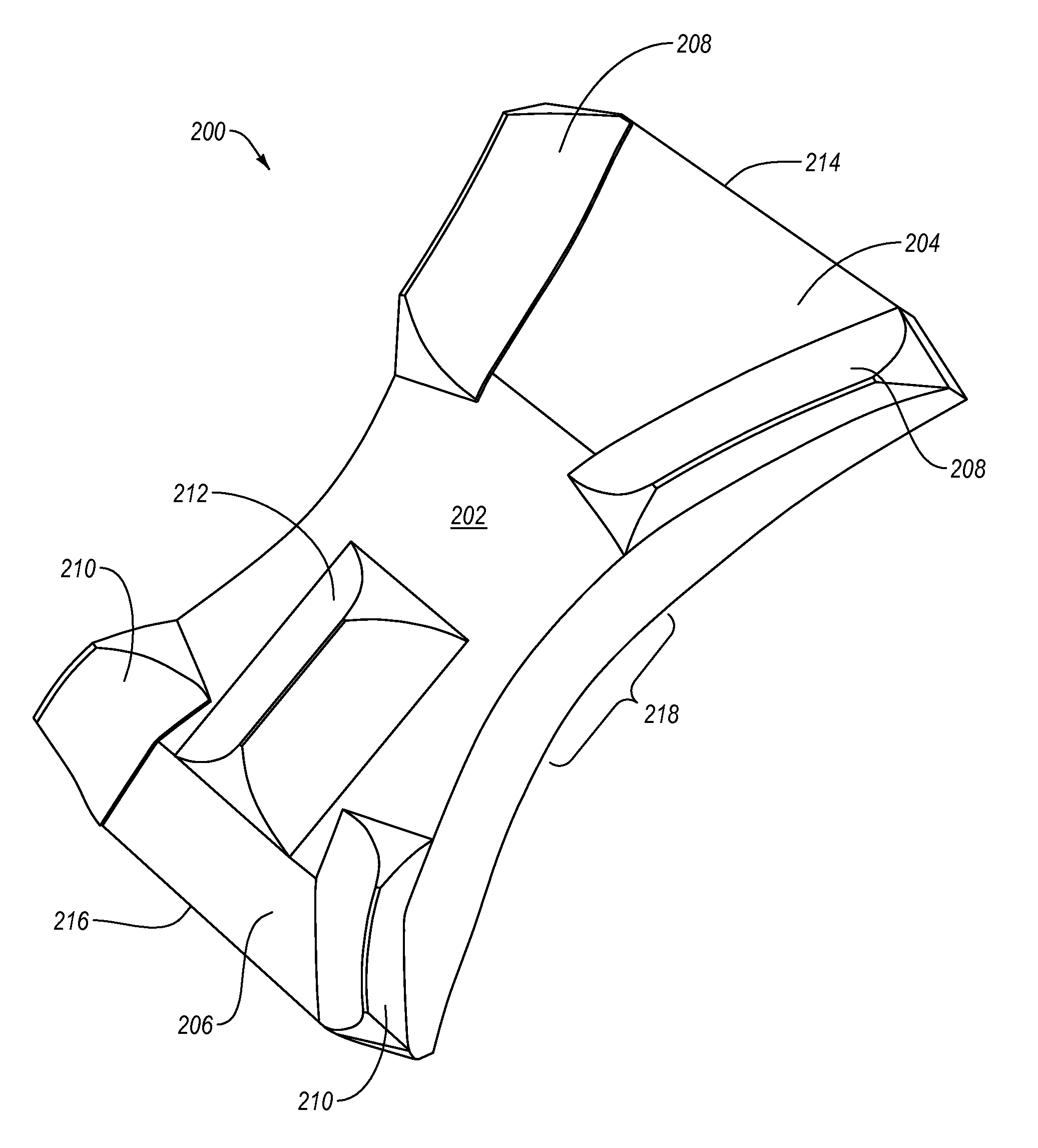
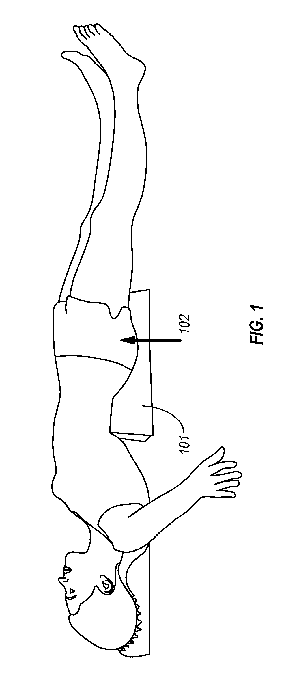
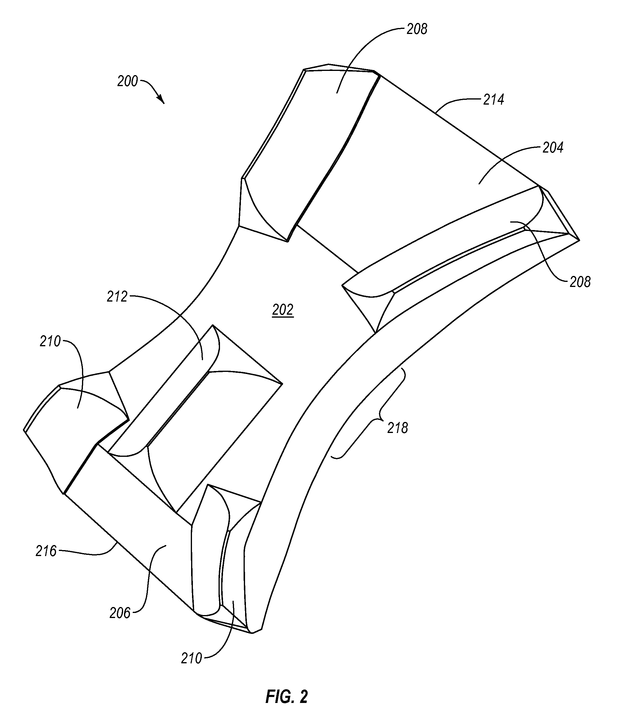
[0021]Implementations of the present invention involve methods and apparatus configured to allow the convenient, reliable and secure positioning of a surgical patient. In one aspect, the invention provides an iliosacral cradle that elevates the ilium and the sacrum, while yet providing stable support to the buttock and flank. The iliosacral cradle is configured to allow for access to the anatomy, e.g., posterolateral buttock for proper positioning of drills, implants, and other surgical equipment to achieve fracture reduction or fixation. The iliosacral cradle elevates the patient's pelvis above the surgical table surface adequately to allow a surgeon to position his / her hands, while holding surgical tools, for the desired posterior-to-anterior vector placement. The cradle distributes the patient's weight over the upper-legs, buttocks, and lower-back of the patient and provides adequate cushioning to hard and soft tissues under pressure. As a result, the cradle provides stable weigh...


PUM


Abstract

Devices and methods for supporting and elevating the pelvis of a patient in a supine position during surgery are disclosed. The device comprises a generally flat main body configured to extend from the patient's lower back to the patient's upper legs, a proximal upper surface with a downward slope towards the proximal end, a distal upper surface with a downward slope towards the distal end, and vertically extending ridges on an upper surface of the device for partially containing one or more portions of the patient's body. In use, the device is positioned on a surgical platform beneath the iliosacral region of the patient.

Description

BACKGROUND OF THE INVENTION[0001]1. The Field of the Invention[0002]The present invention relates to devices and methods for elevating the pelvic position of a surgical patient in a supine position.[0003]2. Background and Relevant Art[0004]Percutaneous bone fixation is a technique used by orthopedic surgeons for the stabilization of unstable fractures. It involves the manipulation, with X-ray guidance, of the fracture into an acceptable position, and the insertion of metal pins through the skin into one bone fragment and across the fracture line into an adjacent bone fragment. Percutaneous fixation falls into the category of minimally invasive surgery, which has become very popular and well described for pelvic and acetabular fractures.[0005]Surgeons commonly perform percutaneous fixation on a radiolucent operating table. A patient undergoing the procedure usually lies in a supine position, i.e. with the front (anterior) facing up and the back (posterior) facing down. Because the op...

Claims


Application Information

Patent Timeline
Patent Type & Authority Patents(United States)
IPC IPC(8): A61G15/00A61G13/12A61G99/00A61F5/01A61B6/04
CPCA61G99/00A61F5/01A61G13/123A61B6/0421A61B6/0442
Inventor ROBRAN, CHAD L.PARSELL, DOUGLAS
Owner BONE FOAM
Why Eureka
Try Eureka