X-ray CT apparatus

A technology of equipment and line data, used in X-ray equipment, medical science, diagnosis, etc., can solve problems such as narrowing of the area, different image quality, and inability to scan

Inactive Publication Date: 2007-09-12
GE MEDICAL SYST GLOBAL TECH CO LLC
View PDF1 Cites 16 Cited by
  • Summary
  • Abstract
  • Description
  • Claims
  • Application Information

AI Technical Summary

Problems solved by technology

As a result, there are wastes and problems: data acquisition has to wait until the carriage velocity on the imaging platform reaches a certain level; requires a start distance until the carriage speed reaches a certain level; There is an area that cannot be scanned in the gantry travel distance of ; and the area that can be scanned is narrowed, or the start of scanning has to wait for the time required to accelerate in the start
However, this causes a problem that the image quality of the tomogram in the constant velocity region of the helical scan differs from that in the acceleration region and the deceleration region

Method used


Image

  • X-ray CT apparatus
  • X-ray CT apparatus
  • X-ray CT apparatus

Examples


Embodiment 1

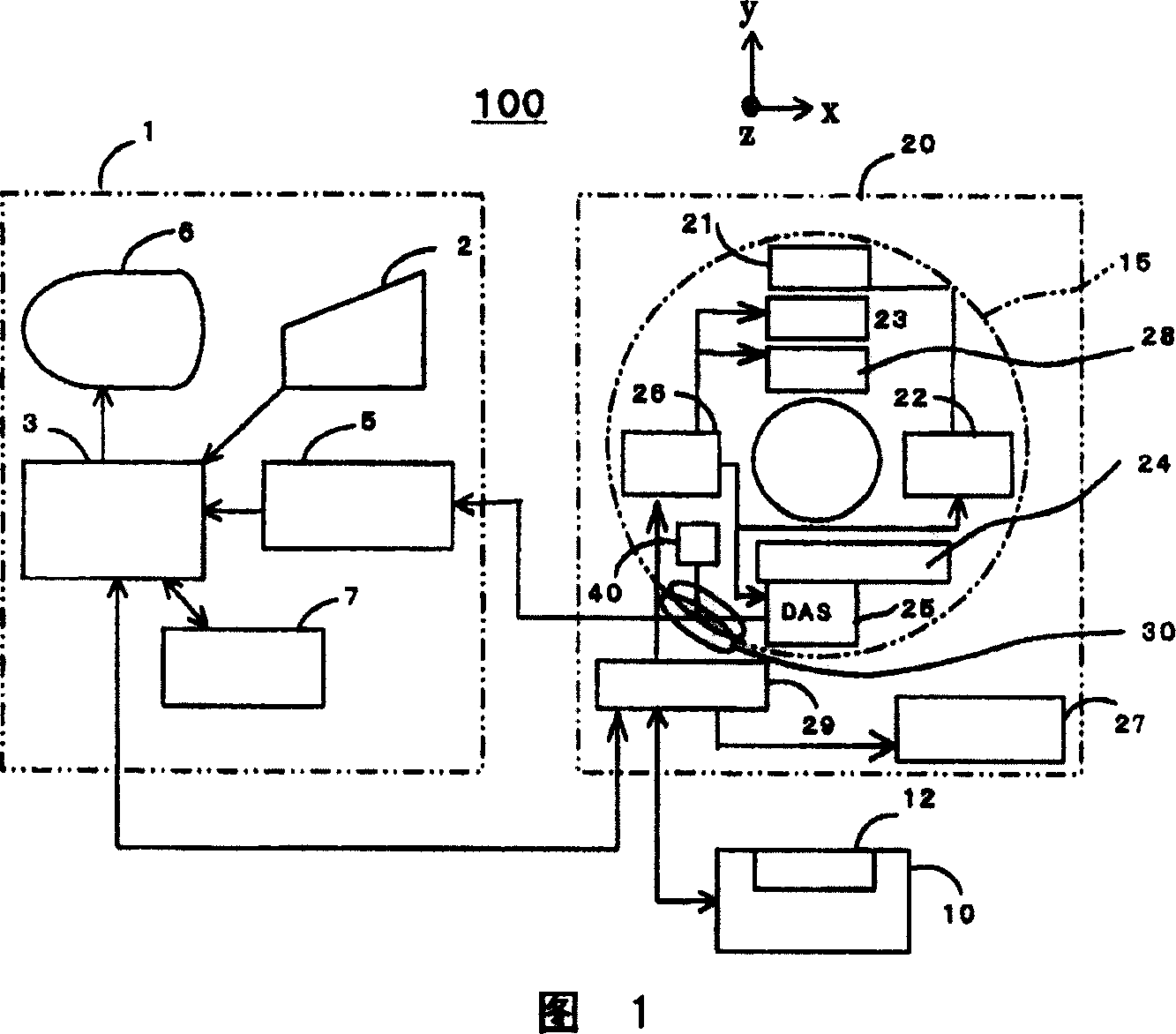
[0134] Embodiment 1: Move the imaging platform 10 or the carriage 12 (hereinafter collectively referred to as the imaging platform 10) in the z direction to collect X-ray data during the acceleration, uniform operation, and deceleration of the imaging platform 10, and after the X-ray data collection is terminated The operation of the imaging platform 10 is ended.

Embodiment 2

[0135] Embodiment 2: Before moving the imaging platform 10 or the carriage 12 (hereinafter collectively referred to as the imaging platform 10) in the z-direction, the imaging platform 10 is kept upright; when X-ray data is collected by conventional scanning (axial scanning), or at fan angle +180 degrees or 360 degrees, or after performing multiple revolutions or film scanning, move the imaging platform 10 to collect X-ray data during the acceleration, uniform operation, and deceleration of the imaging platform 10; after stopping the operation of the imaging platform 10, when imaging The platform 10 is at the standing time, and performs traditional scanning (axial scanning) or cine scanning to collect X-ray data at the fan angle +180 degrees or 360 degrees, or multiple revolutions; after the X-ray data collection is completed, the X-ray data is also terminated. Radiation.

[0136] Example 1

[0137] FIG. 20 shows a flow chart of the entire operation flow of the first embodiment.

...

Embodiment 3

[0367] In Embodiment 1 and Embodiment 2, the z coordinate at each time point is predicted as shown in the graph of FIG. 21 or 23. Or use an encoder or the like arranged on the imaging platform 10 or the bracket 12 to measure the z-direction coordinate position, and extract the X-ray projection data of FIG. 10 when the three-dimensional image is reconstructed for measuring each angle of view or a plurality of fixed intervals. In the case of the z-direction coordinate position of the viewing angle, accurate three-dimensional back projection can be realized in consideration of the z-direction coordinate positions of each viewing angle or multiple viewing angles at fixed intervals calculated based on these predicted or measured viewing angles.

[0368] This makes it possible to obtain a tomogram with high picture quality, uniform picture quality in the z-direction, and relatively free of artifacts.


PUM


Abstract

X-ray CT apparatus includes an x-ray data collecting device for collecting x-ray projection data transmitted by a subject positioned between an x-ray generating device and a multi-row x-ray detector, while rotating said x-ray generating device and said multi-row x-ray detector around a rotation center positioned in-between, an image reconstructing device for performing image reconstruction from the projection data collected from the x-ray data collecting device, an image display device for displaying a tomogram obtained by image reconstruction, and a scanning condition setting device for setting various scanning conditions of tomography scanning. The x-ray data collecting device is operable for variable-pitch helical scanning which x-ray projection data of the subject on a scanning table is collected by moving the scanning table while varying the speed relative to a scanning gantry in a z direction perendicular to an xy plane which is the rotating plane of the x-ray generating device and the two-dimensional x-ray area detector, and of which starting of the x-ray data collection and starting of the scanning table movement relative to the scanning gantry and / or stopping of the x-ray data collection and stopping of the scanning table movement relative to the scanning gantry are asynchronously executed.

Description

Technical field [0001] The present invention relates to a medical X-ray CT (computed tomography) device or industrial X-ray CT equipment that can improve the image quality of an imaging method. Background technique [0002] Conventionally, in X-ray CT equipment using multiple rows of X-ray detectors or two-dimensional X-ray area detectors represented by flat-panel X-ray detectors, the uniform spiral scanning is performed at a uniform speed. The data collected in the part is shown in Fig. 16 (see, for example, JP-A No. 2004-073360). As a result, there is such a waste and problem: data collection has to wait until the speed of the carriage on the imaging platform reaches a certain level; it needs to move the distance together until the speed of the carriage reaches a certain level; accordingly, it is at the same length as the starting distance. There is an area that cannot be scanned in the travel distance of the gantry; and the area that can be scanned is narrowed, or the start of...

Claims


Application Information

Patent Timeline
Patent Type & Authority Applications(China)
IPC IPC(8): A61B6/03G01N23/04H05G1/02
CPCA61B6/469A61B6/032A61B6/04A61B6/507A61B6/467A61B6/4085A61B6/027A61B6/03
Inventor 西出明彦萩原明森川琴子
Owner GE MEDICAL SYST GLOBAL TECH CO LLC
Why Eureka
Try Eureka