Planar angular visualization of the bronchial tree

A bronchial tree and plane angle technology, applied in the field of medical imaging, can solve the problems of difficult observation of the airway passage and time-consuming visual inspection, and achieve the effect of convenient automatic or manual processing and easy visual inspection

Active Publication Date: 2007-10-31
KONINKLIJKE PHILIPS ELECTRONICS NV
View PDF0 Cites 4 Cited by
  • Summary
  • Abstract
  • Description
  • Claims
  • Application Information

AI Technical Summary

Problems solved by technology

Therefore, visual inspection of all pathways in the 3D bronchial tree is time consuming
On the other hand, the planar projection of the three-dimensional tree lead

Method used


Image

  • Planar angular visualization of the bronchial tree
  • Planar angular visualization of the bronchial tree
  • Planar angular visualization of the bronchial tree

Examples


Embodiment Construction

[0023] The following description focuses on one embodiment of the invention, applicable to computed tomography (CT) systems, and in particular to multi-slice CT data. However, it should be understood that the invention is not limited to this application, but may be applied to many other imaging systems including, for example, Magnetic Resonance Imaging (MRI) systems, Three-dimensional Rotational Radiography (3D-RA) scanners, Position Emission Tomography (PET ) scanners, single photon emission computed tomography (SPECT) scanners, and the like.

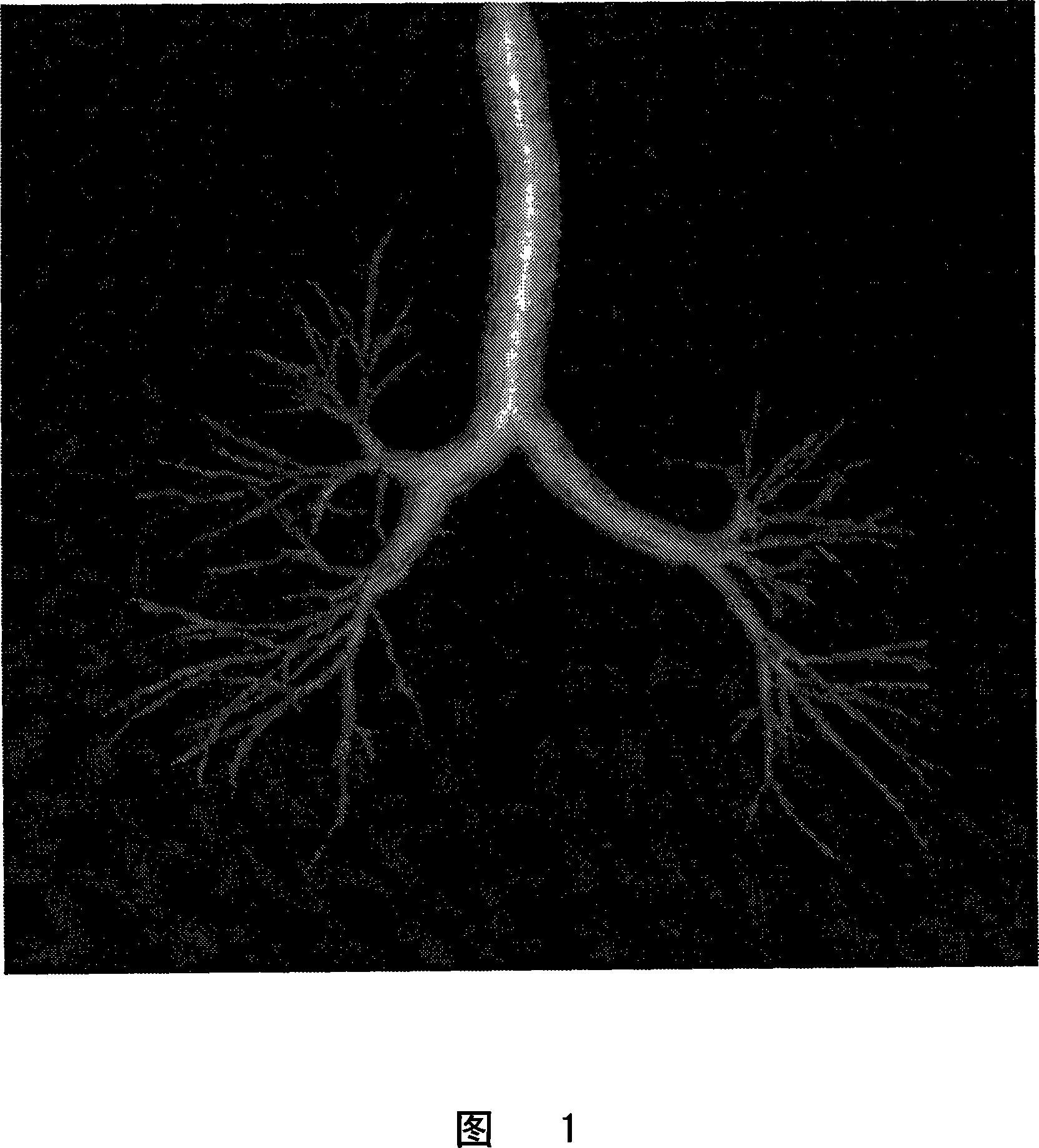
[0024] In the following, an automated method for extracting anatomical trees from multi-slice CT data is described. While much of the discussion of the present invention will refer to bronchial trees, those skilled in the art will understand that the present invention relates to any type of anatomical tree, such as a vascular tree, and the invention is not limited thereto.

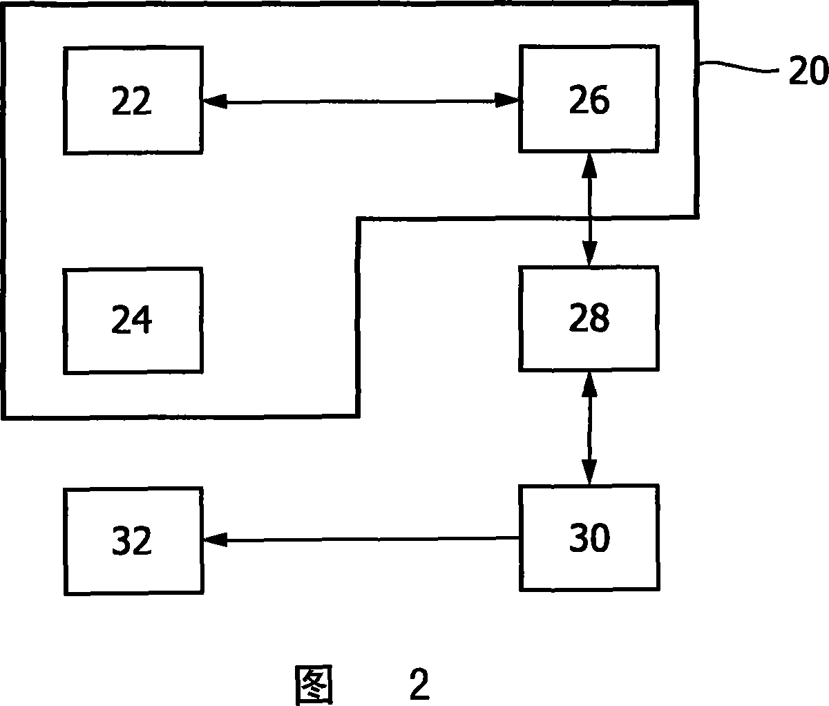
[0025] An example of a CT acquisition device 20 according to...


PUM


Abstract

A system (20) an image aquisition device, an image workstation, computer- readable medium and method for visualizing an anatomical tree structure is disclosed. The anatomical tree structure is segmented from a three-dimensional image set of at least a portion of a body. A planar angular visualization of the anatomical tree structure is then determined. The planar angular visualization of the anatomical tree structure is then displayed. Since the displayed anatomical tree structure has no occlusions or intersections of segments, the anatomical tree is much easier to read which facilitates subsequent automated or manual diagnosis of the tree for anomalies.

Description

technical field [0001] The present invention relates generally to the field of medical imaging. More particularly the present invention relates to methods and apparatus for planar angular visualization of anatomical tree structures, and more particularly to planar topological mapping of the three-dimensional bronchial tree structure of the lung . Background technique [0002] In helical computed tomography (CT) imaging, the patient is advanced linearly through the examination area in a direction perpendicular to the plane of gantry rotation to effect a helical rotational motion of the X-ray source around the object. X-ray absorption data acquired during the helical rotational motion is reconstructed using filtered back-projection or another reconstruction method to generate a three-dimensional image representation of the object, or a selected portion thereof. [0003] High resolution CT datasets from multi-slice scanners allow inspection of the bronchi down to smaller airw...

Claims


Application Information

Patent Timeline
IPC IPC(8): G06T5/00G06T11/00
CPCG06T7/0081G06T2207/10081G06T2207/30101G06T7/11
Inventor R·韦姆克T·布拉弗特T·比劳
Owner KONINKLIJKE PHILIPS ELECTRONICS NV
Why Eureka
Try Eureka