Biomarkers for breast cancer

A biomarker, technology for breast cancer, applied in the field of clinical diagnostics

Inactive Publication Date: 2008-07-16
THE JOHN HOPKINS UNIV SCHOOL OF MEDICINE
View PDF18 Cites 3 Cited by
  • Summary
  • Abstract
  • Description
  • Claims
  • Application Information

AI Technical Summary

Problems solved by technology

Currently, no molecular biomarker is recommended for early detection of breast cancer (Smith, R.A. et al. (2004) CA Cancer J. Clin. 54: 41-52)

Method used


Image

  • Biomarkers for breast cancer
  • Biomarkers for breast cancer
  • Biomarkers for breast cancer

Examples


Embodiment 1

[0170] 11.1. Example 1. Discovery of breast cancer biomarkers

[0171] It is generally understood that the embodiments and specific embodiments described herein are for illustrative purposes only, and for those skilled in the art, various modifications or changes according to these embodiments are also considered to have been suggested by the present case and included in the within the spirit and scope of the invention and within the purview of the appended claims. All publications, patents, and patent applications cited herein are hereby incorporated by reference for all purposes.

Embodiment 2

[0172] 11.2. Example 2. Material Level Methods

[0173] 11.2.1. Multi-center study design

[0174] To minimize potential bias in patient selection and fluid collection procedures at each institution, and to maximize the applicability of this finding, we selected three institutions: University of Texas M.D. Anderson Cancer Center; Sidney Kimmel Comprehensive Cancer Center in Johns Hopkins Hospital; And Department of Surgery, Feinberg School of Medicine, Northwestern University enlisted two liquid samples for nipple aspiration and milk duct lavage. Sample collection at each site was approved by the respective Institutional Review Board, and current proteosome studies were approved by the Institutional Review Board of Johns Hopkins University.

[0175] 11.2.2. Patient - Nipple Aspiration Fluid (NAF)

[0176] NAF samples were obtained from the University of Texas M.D. Anderson Cancer Center. Patients eligible for bilateral nipple aspiration were those with biopsy-proven stage I...

Embodiment 3

[0198] 11.3. Example 3. Results

[0199] 11.3.1. Proteome profile of breast fluid

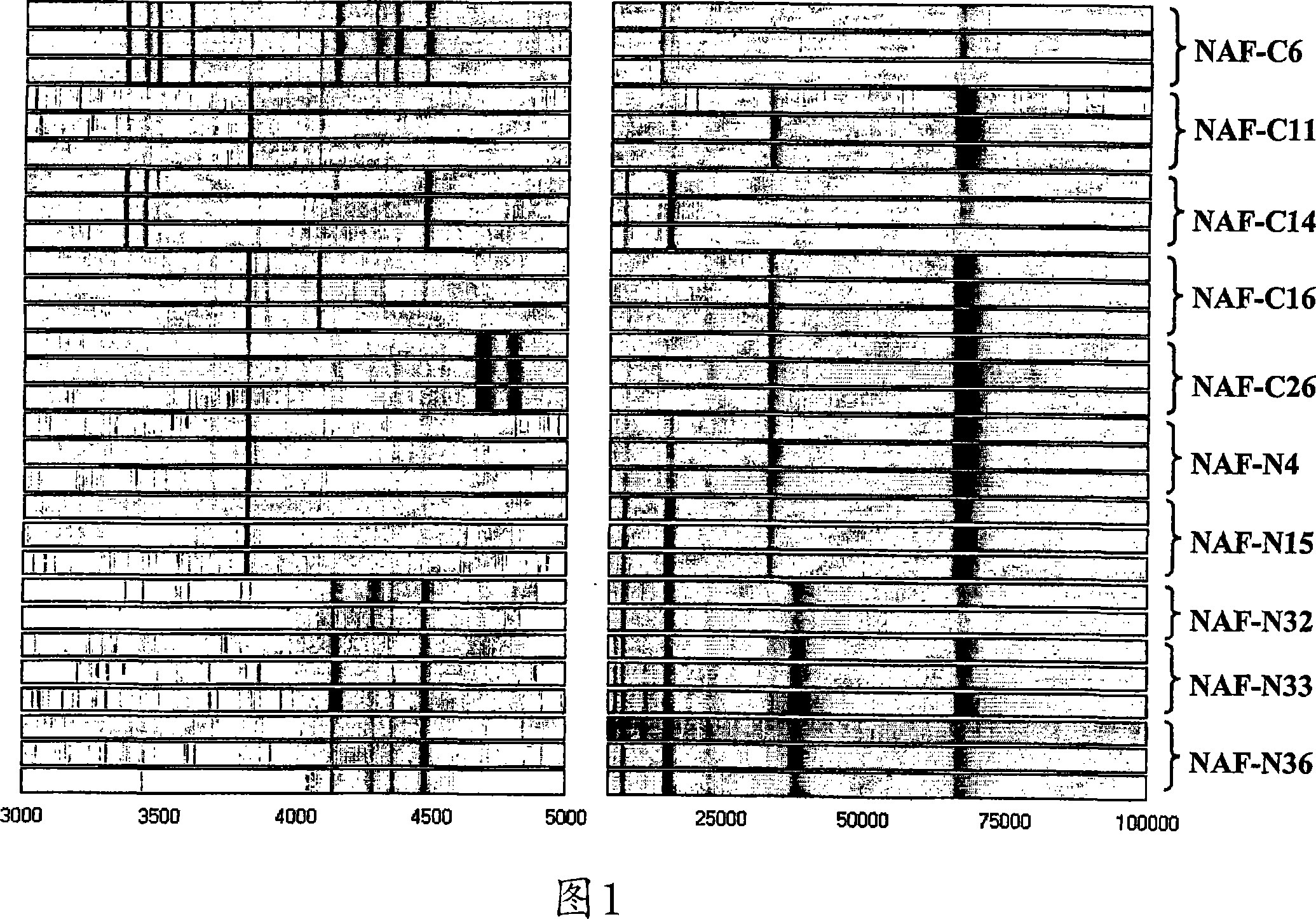
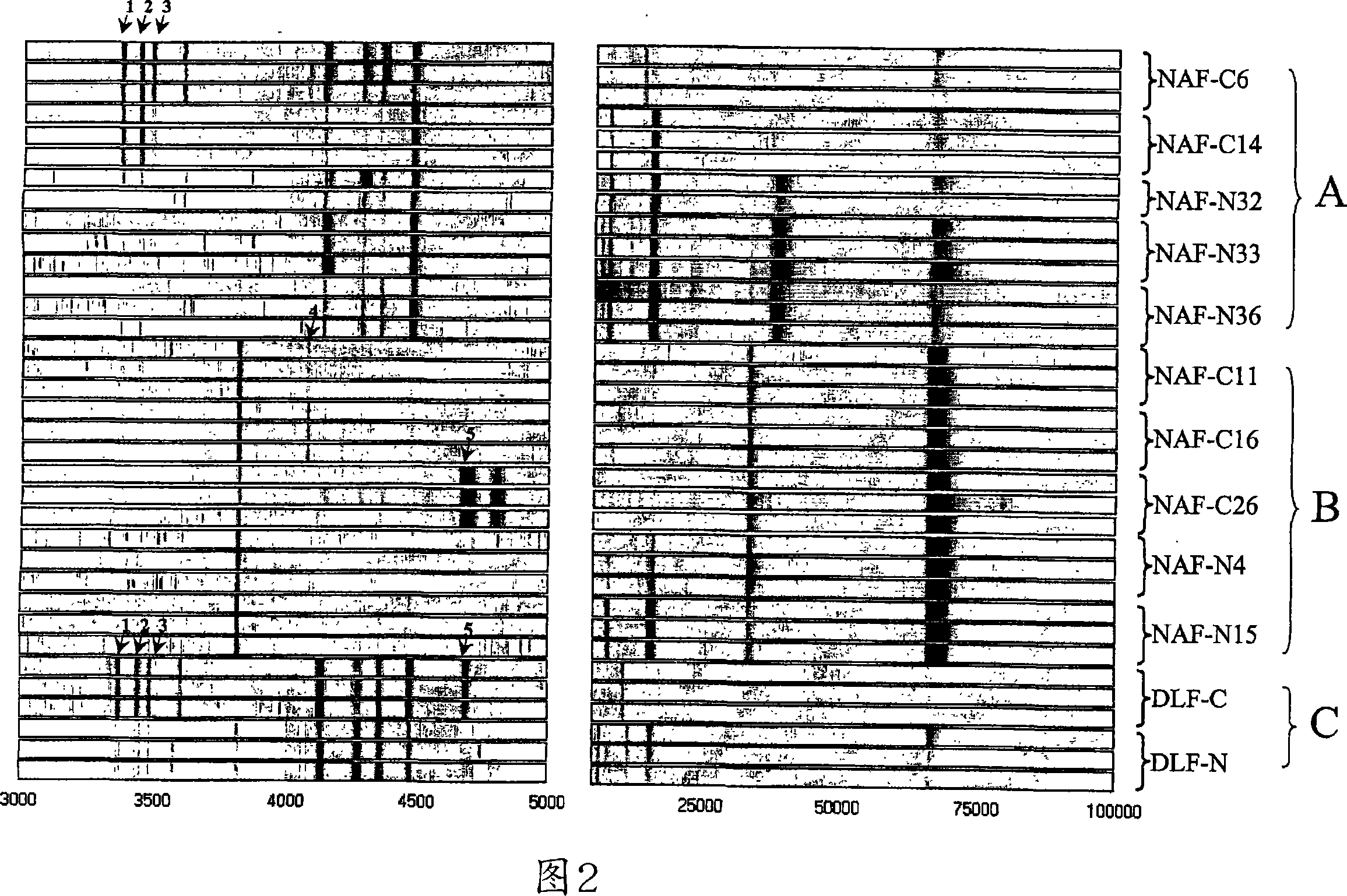
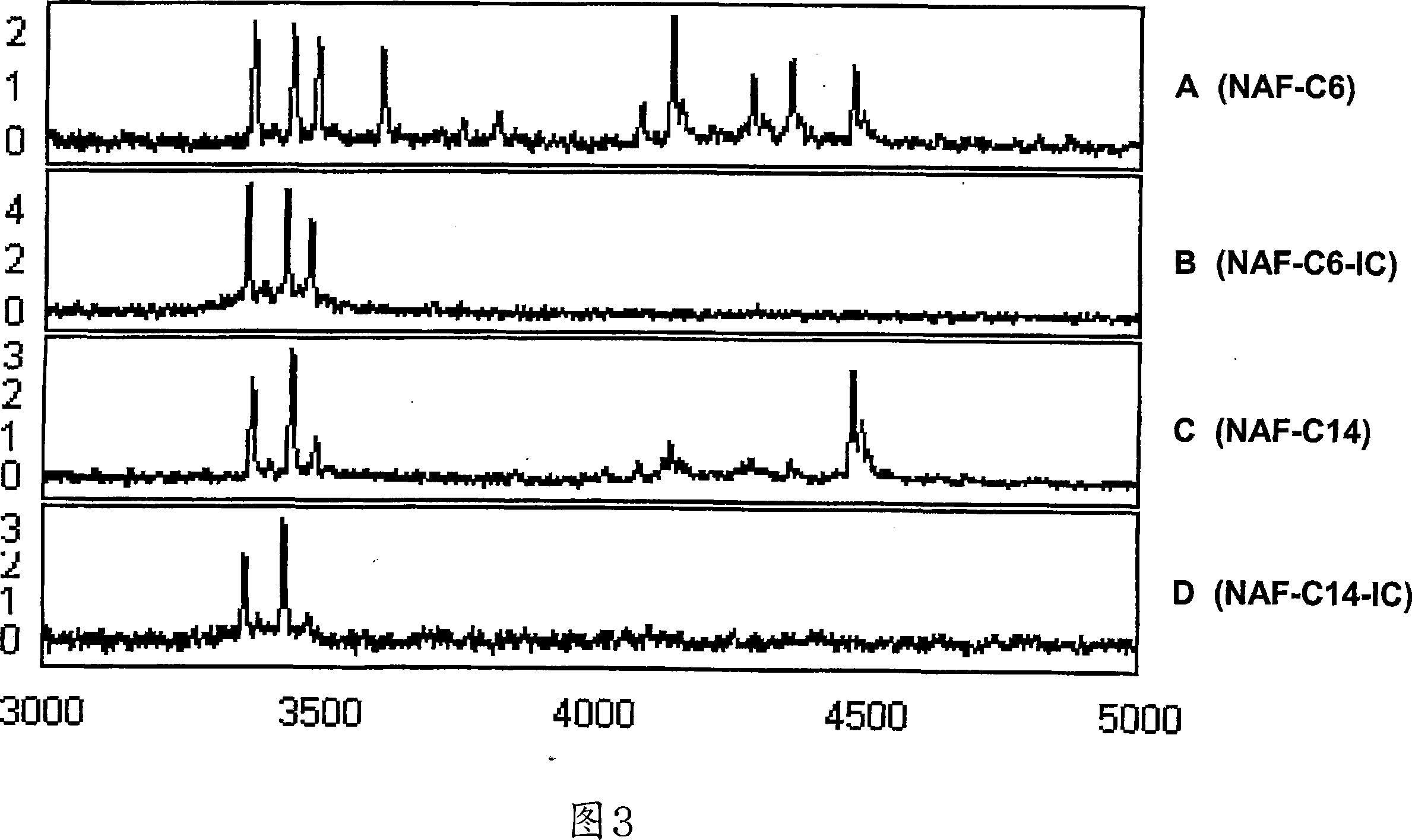
[0200] Reproducible protein profiles were obtained using our optimized microarray procedure and breast fluid samples containing 1 μg of whole protein. Figure 1 shows nipple suction fluid from 5 cancerous breasts (C6, C11, C14, C16, C26) of patients with primary invasive cancer and 5 normal control breasts (N4, N15, N32, N33, N36) Pseudo-gel map of the protein map. The general protein expression profile varies from subject to subject, whereas mass spectra from triplicate analyzes of the same specimen are highly reproducible. M / Z (mass / charge) ion signals less than 3000 are mainly matrix material noise, and a maximum M / Z is detected at 135,000. We have manually selected 73 protein peaks (signal / noise >5, M / Z from 3K to 135K) for subsequent biomarker evaluation.

[0201] 11.3.2. Using training data to select biomarkers

[0202] Use 10 NAF samples as training samples. First, we performed an u...


PUM


Abstract

The present invention provides protein-based biomarkers and biomarker combinations that are useful in qualifying breast cancer status in a patient. In particular, the biomarkers of this invention are useful to classify a subject sample as breast cancer or non-breast cancer. The biomarkers can be detected by SELDI mass spectrometry.

Description

[0001] Related applications [0002] This application claims priority to US Provisional Application 60 / 685,459, filed May 26, 2005, which is hereby expressly incorporated by reference in its entirety. 【Technical field】 [0003] In general, the present invention pertains to clinical diagnostics. 【Background technique】 [0004] Breast cancer is the most commonly diagnosed cancer in women. Presymptomatic screening to detect early-stage breast cancer while it is still resectable and has a chance of cure can greatly reduce breast cancer-related mortality. Unfortunately, only 63% of breast cancers (1992-1999, USA) are detected at diagnosis (Jemal, A. et al. (2004) CA Cancer J. Clin. 54:8-29). Small lesions are often missed or not seen even with mammography, especially in young women and women with dense breast tissue (Antman, K. and Shea, S. (1999) JAMA 281: 1470-1472). The potential to identify molecular markers of these small lesions that are invisible to imaging techniques w...

Claims


Application Information

Patent Timeline
Patent Type & Authority Applications(China)
IPC IPC(8): G01N33/574G06F19/00C07K5/00A61K39/00
CPCG01N2333/4721G01N33/57415
Inventor J·李S·苏库马尔D·W·尚
Owner THE JOHN HOPKINS UNIV SCHOOL OF MEDICINE
Why Eureka
Try Eureka