
Laser treatment apparatus
A technology of laser therapy and laser beams, which is applied in the direction of cardiac catheterization, catheterization, application, etc., and can solve problems such as not disclosing specific structures
- Summary
- Abstract
- Description
- Claims
- Application Information
AI Technical Summary
Problems solved by technology
Method used
Image

Examples
no. 1 approach
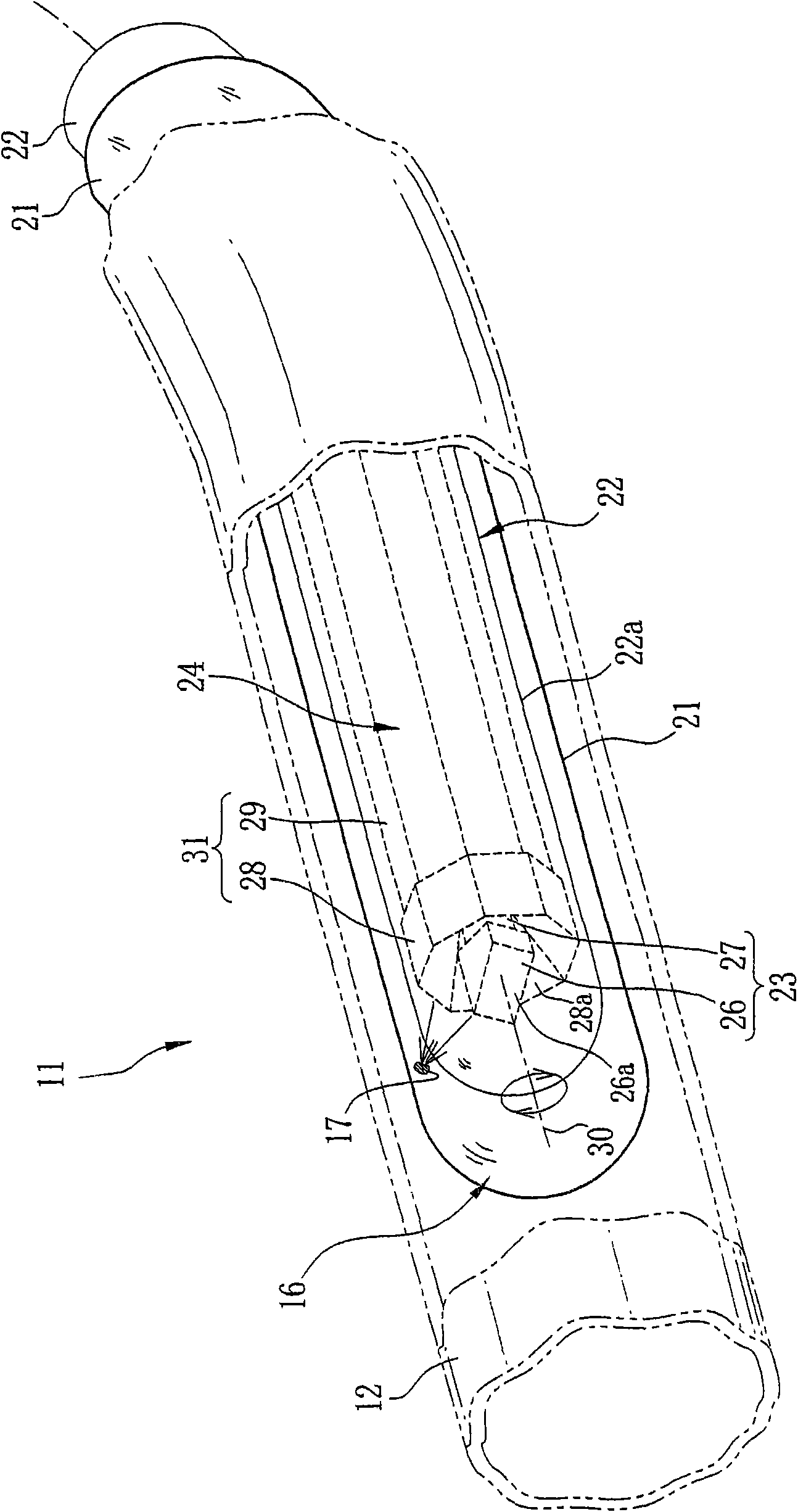
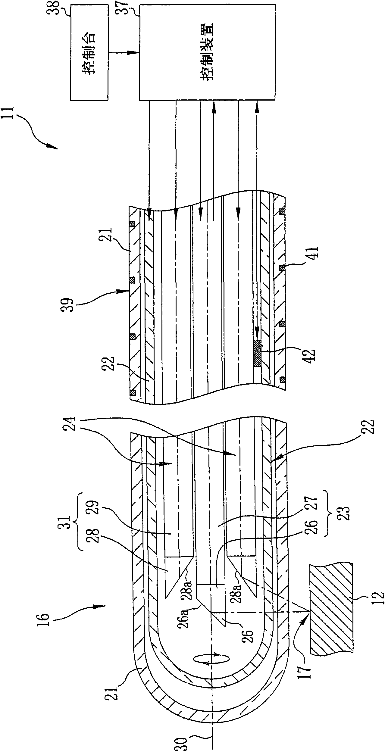
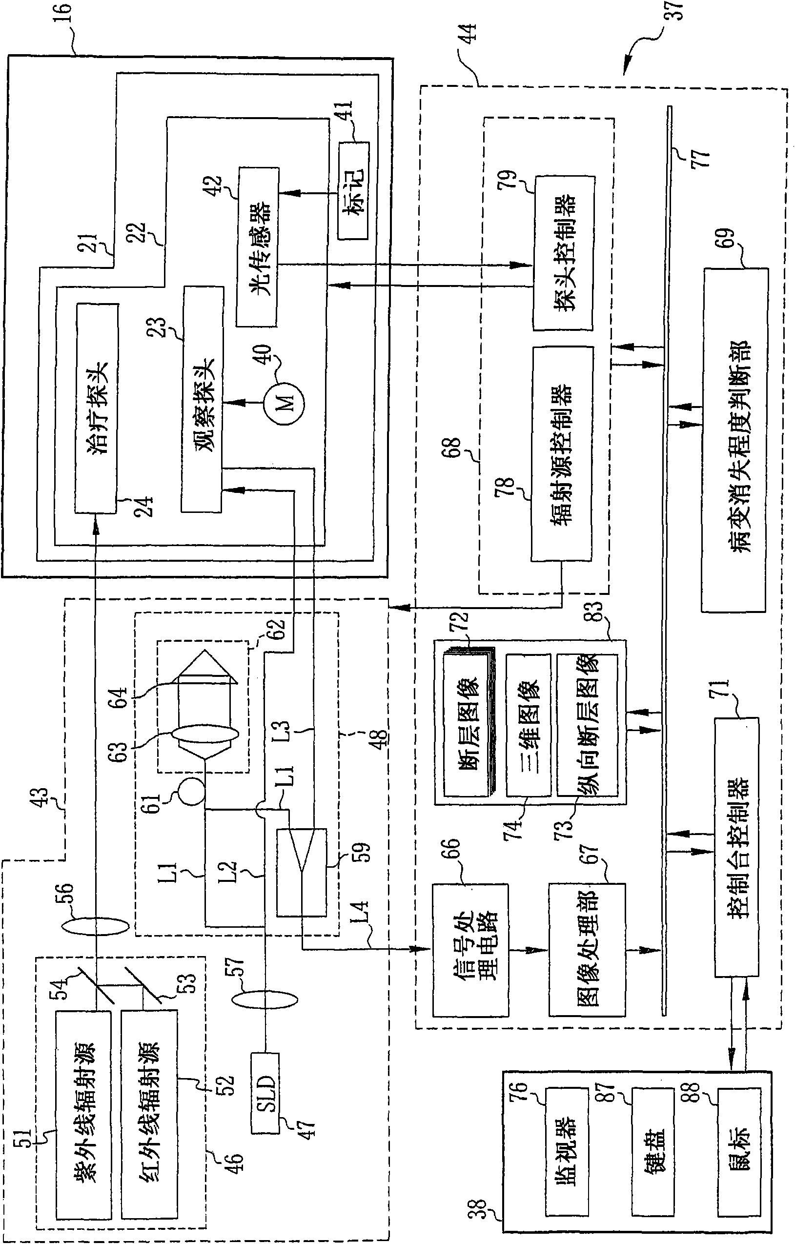
[0081] In the following first to third embodiments, the laser treatment device 11 is set in the auxiliary mode. In the first embodiment, the degree of disappearance of the lesion 17 is judged every time the laser beam has been applied for a predetermined time, and the laser source portion 46 is turned on and off in response to the judgment result. When the catheter assembly 16 is introduced into the blood vessel 12 , the probe unit 22 sweeps over, and a tomographic image 72 of the blood vessel 12 is generated from the signal of the observation probe 23 .
[0082] The tomographic image 72 is displayed on the monitor 76 in real time, such as Figure 4 shown in . A parallel tomographic image 73 and a 3D image 74 are generated from the obtained plurality of tomographic images 72 . The parallel tomographic image 73 selected by the console 38 ( Figure 4 left part of ) and 3D image 74 ( Figure 4 The right part of ) is displayed on the screen of the monitor 76 together with the ...
no. 2 approach
[0097] In a second embodiment, the power level of the laser beam is controlled based on the degree of disappearance of the lesion 17 . As in the first embodiment, an observation image is obtained (step S1), and the brightness value of the lesion 17 is specified (step S2). Then, a treatment area is designated in the blood vessel axial direction of the blood vessel 12 (step S3), and laser irradiation conditions are set (step S4). When the laser radiation operation is performed (step S5), the laser treatment apparatus 11 judges whether or not there is a pixel having a specified brightness value in the laser tomographic image 72 (step S6).
[0098] As a result of the judgment, when the lesion 17 exists in the tomographic image 72 , the lesion disappearance degree judging section 69 judges the thickness of the lesion 17 in the designated laser emission direction, and inputs the thickness into the CPU 68 . The CPU 68 compares the thickness of the lesion 17 with a predetermined thre...
PUM
 Abstract
Description
Claims
Application Information