Quantification analisys of immobilized contrast agent in medical imaging applications

A contrast agent, circulating function technology, used in the field of medical imaging, to solve problems such as blocking, detecting and quantifying fixed contrast agents

Active Publication Date: 2010-12-15
BRACCO SUISSE SA
View PDF7 Cites 13 Cited by
  • Summary
  • Abstract
  • Description
  • Claims
  • Application Information

AI Technical Summary

Problems solved by technology

Therefore, fixed contrast agents cannot be detected and quantified with acceptable accuracy
This prevents the clinical application of known analytical techniques based on the use of targeted contrast agents

Method used


Image

  • Quantification analisys of immobilized contrast agent in medical imaging applications
  • Quantification analisys of immobilized contrast agent in medical imaging applications
  • Quantification analisys of immobilized contrast agent in medical imaging applications

Examples


Embodiment Construction

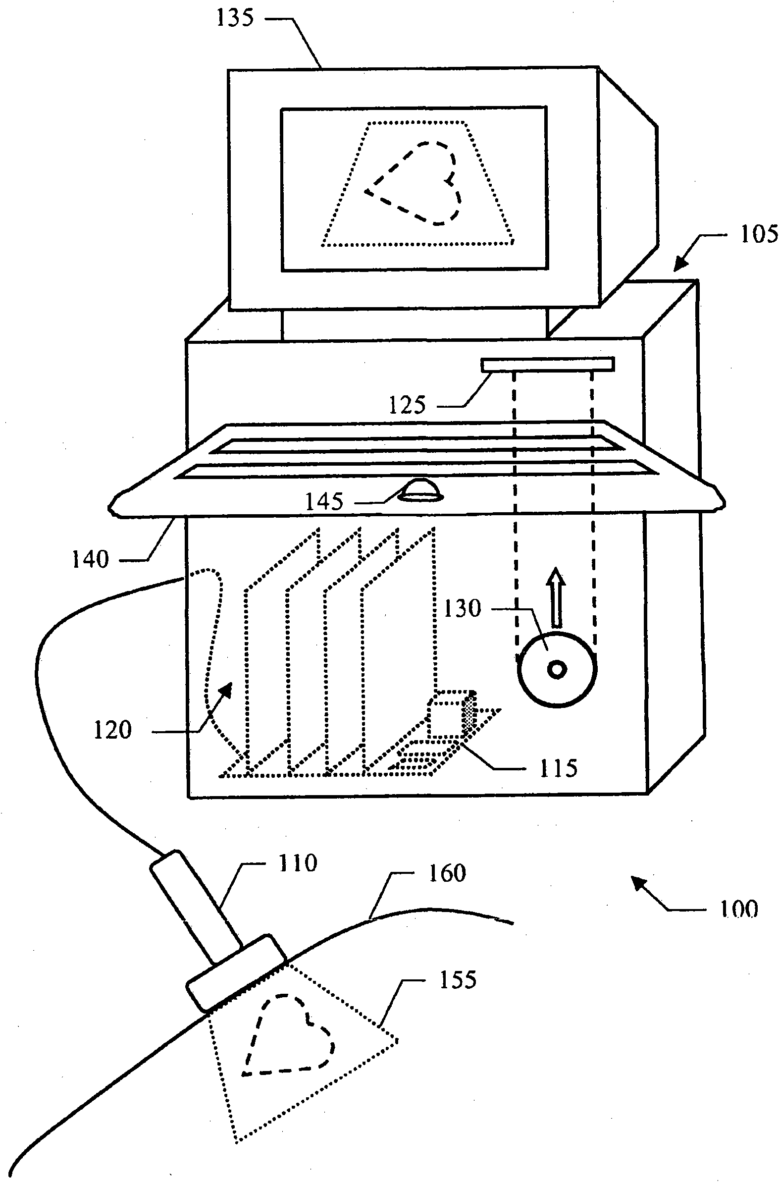
[0036] In particular, refer to figure 1, shows a medical imaging system including an ultrasound scanner 100 . The ultrasound scanner 100 includes a central unit 105 and a handheld transmit-receive imaging probe 110 (eg, of the array type). Imaging probe 110 transmits ultrasound waves comprising a train of pulses (e.g., with a center frequency between 1 and 50 MHz) and receives raw radio frequency (RF) echo signals obtained from reflections of the ultrasound pulses; The pulse-echo mode uses the transmit / receive multiplexer of the imaging probe 110

[0037] The central unit 105 houses a motherboard 115 on which are mounted electronic circuits that control the operation of the ultrasound scanner 100 (eg, a microprocessor, working memory, and a hard drive). Additionally, one or more daughter boards (generally indicated at 120 ) are plugged onto motherboard 115 ; daughter boards 120 provide electronics for driving imaging probe 110 and for processing received echo signals. The u...


PUM


Abstract

A solution is proposed for analyzing a body part (155) of a patient (160), which is perfused with a contrast agent capable of circulating within the patient and of being substantially immobilized on a biological target. A corresponding system (105) includes means (405-440) for providing an echo signal indicative of a response over time to an interrogation signal of the body part, means (442,450) for associating the echo signal with a model function of time modeling an evolution over time of the contrast agent in the body part, the model function including a combination of a circulation function modeling the circulation of the contrast agent and a decay function modeling a decay of the echo signal from the immobilized contrast agent, and means (455-475) for estimating at least one kinetics indicator of the contrast agent from the model function.

Description

technical field [0001] The solutions according to the embodiments of the present invention relate to the field of medical imaging. More specifically, this approach relates to medical imaging applications based on contrast agents adapted to be immobilized on biological targets. Background technique [0002] Medical imaging is a well-established technique (in the field of medically applied equipment) that allows analysis of body parts of a patient in a substantially non-invasive manner. Certain medical imaging techniques are based on administering to the patient an ultrasound contrast agent (UCA) (e.g., a suspension comprising phospholipid-stabilized gas-filled microbubbles); The contrast agent can be easily detected. The echo signals thus obtained can be used to estimate different physiological properties of the body part; for example, when the contrast agent flows at the same speed as the patient's blood, its tracking provides an indication of the perfusion of blood in the...

Claims


Application Information

Patent Timeline
Patent Type & Authority Applications(China)
IPC IPC(8): A61B8/00G16B5/30
CPCA61B8/481G16B5/00G16B5/30A61B8/14A61B8/5223
Inventor M·阿迪特T·梅萨格M·施奈德P·弗林京
Owner BRACCO SUISSE SA
Why Eureka
Try Eureka