
Ultrasonic diagnostic apparatus, method for controlling display of image and control program of the same

A technology of ultrasonic diagnosis and equipment, which is applied in the directions of ultrasonic/sonic wave/infrasonic wave diagnosis, sound wave diagnosis, infrasonic wave diagnosis, etc., and can solve the problem that the elasticity of living tissue is not accurately grasped

Inactive Publication Date: 2012-01-25
GE MEDICAL SYST GLOBAL TECH CO LLC
View PDF11 Cites 9 Cited by
  • Summary
  • Abstract
  • Description
  • Claims
  • Application Information

AI Technical Summary

Problems solved by technology

Thus, there may be a possibility that the elasticity of living tissue is not accurately grasped

Method used


Image

  • Ultrasonic diagnostic apparatus, method for controlling display of image and control program of the same
  • Ultrasonic diagnostic apparatus, method for controlling display of image and control program of the same
  • Ultrasonic diagnostic apparatus, method for controlling display of image and control program of the same

Examples


no. 1 example

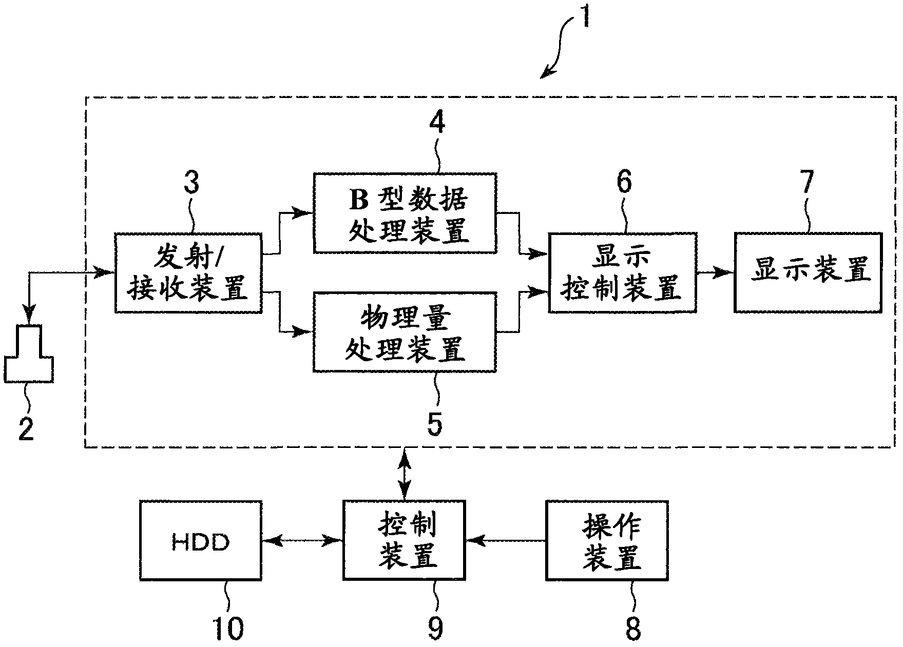
[0046] First, the first embodiment will be based on Figure 1 to Figure 13 illustrate. exist figure 1 The ultrasonic diagnostic equipment 1 shown in includes an ultrasonic probe 2, a transmitting / receiving device 3, a B-type data processing device 4, a physical quantity processing device 5, a display control device 6, a display device 7, an operating device 8, a control device 9 and an HDD ( hard drive)10.

[0047] The ultrasonic probe 2 transmits ultrasonic waves to living tissues and receives echo signals. Under the condition that the ultrasonic probe 2 is in contact with the surface of the living tissue, the pressing and releasing motion of the ultrasonic probe 2 is repeated and the ultrasonic probe 2 applies acoustic radiation pressure to the living tissue. As a result, the ultrasonic probe 2 acquires echo data as transmission / reception of ultrasonic waves is performed, deforming living tissue. Based on this acquired echo data, an elasticity image is generated as desc...


PUM


Abstract

An ultrasonic diagnostic apparatus includes a physical amount calculation device configured to calculate a physical amount on elasticity of living tissue based on an echo signal obtained by transmitting an ultrasonic wave to the living tissue, and a display image control device configured to control display of a predetermined alternative elastic image displayed instead of an elastic image based on the physical amount calculated for error frames using a ratio of non-error frames or the error frames in a predetermined plurality of frames, wherein the error frames are determined as not meeting a standard.

Description

technical field [0001] The present invention relates to an ultrasonic diagnostic apparatus and more particularly to an apparatus for displaying an elasticity image indicating the hardness or softness of living tissue and a control program thereof. Background technique [0002] For example, an ultrasonic diagnostic apparatus for displaying a combined image of a normal B-mode image and an elastic image indicating the hardness or softness of living tissue is disclosed in Japanese Examined Patent No. 3932482 (US Publication No. 20060052702A1). In this type of ultrasonic diagnostic apparatus, an elastic image is produced as follows. First, transmission / reception of ultrasonic waves is performed to obtain echo signals by repeating pressing and releasing motions with an ultrasonic probe. Then, based on the obtained echo data, a physical quantity of elasticity of living tissue is calculated, and the obtained physical quantity is converted into tone information to generate a color e...

Claims


Application Information

Patent Timeline
Patent Type & Authority Applications(China)
IPC IPC(8): A61B8/00
CPCA61B8/485G01S7/52034G01S7/5206A61B8/5269A61B8/08G01S7/52042A61B8/14G16Z99/00
Inventor 谷川俊一郎
Owner GE MEDICAL SYST GLOBAL TECH CO LLC
Features
  • Generate Ideas
  • Intellectual Property
  • Life Sciences
  • Materials
  • Tech Scout
  • Unparalleled Data Quality
  • Higher Quality Content
  • 60% Fewer Hallucinations
Learn More