Image postprocessing method for removing metal artifact from computed tomography (CT) image

A technology for metal artifacts and CT images, applied in image data processing, image enhancement, instruments, etc., can solve problems such as difficulties, artifacts that have not been removed, and problems that cannot be used for clinical diagnosis

Active Publication Date: 2012-07-11
PHILIPS CHINA INVESTMENT
View PDF3 Cites 29 Cited by
  • Summary
  • Abstract
  • Description
  • Claims
  • Application Information

AI Technical Summary

Problems solved by technology

[0014] (2) For the multi-slice CT using the 3D reconstruction algorithm, this method needs to process a large amount of projection data, and it faces great difficulties in terms of processing effect and processing efficiency
[0020] (1) The better method in image domain processing is t

Method used


Image

  • Image postprocessing method for removing metal artifact from computed tomography (CT) image
  • Image postprocessing method for removing metal artifact from computed tomography (CT) image
  • Image postprocessing method for removing metal artifact from computed tomography (CT) image

Examples


Embodiment Construction

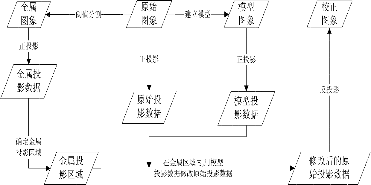
[0098] The present invention removes the image postprocessing method of metal artifact in the CT image comprising the following steps:

[0099] Convert the original CT image from a rectangular coordinate image to a polar coordinate image; determine the metal projection area in the polar coordinate image; build a model in the polar coordinate image; use the above model for model correction; correct the positive and negative introduced in the above model correction Projection error; transforms a polar coordinate image into a rectangular coordinate image.

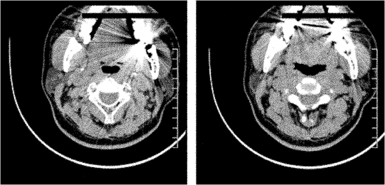
[0100] The method of the present invention is a method for image post-processing, that is, the reconstructed image (called the original image) with metal artifacts is processed, and its basic idea is as follows: firstly, the original image is forward-projected to obtain the original image's forward projection data (referred to as original projection data); then the original image is thresholded, segmented into metal images, an...


PUM


Abstract

The invention relates to an image postprocessing method for removing a metal artifact from a computed tomography (CT) image, which comprises the following steps of: converting an original CT image into a polar coordinate image from an orthogonal coordinate image; determining a metal projection region in the polar coordinate image; establishing a model in the polar coordinate image; performing model correction by adopting the model; correcting front and back projection errors brought into the model correction; and converting the polar coordinate image into the orthogonal coordinate image. According to a model establishing method provided by the invention, the good model can be established, and finally, a good correction result can be obtained; the errors brought in by front and back projection are corrected, so that the result after the correction is better, and the processing speed is ensured.

Description

technical field [0001] The invention relates to a CT image processing technology in the medical field, in particular to an image post-processing method for removing metal artifacts in a CT image. Background technique [0002] Clinically, if the scanned fault contains metal, due to the high attenuation characteristics of the metal to X-rays, the corresponding channel value passing through the metal has serious measurement errors (this paper refers to such a channel as a damaged channel). Make the reconstructed image produce something like figure 1 Artifacts are shown, which appear primarily as black and white streaks emanating from the metal and radial lines that fill the image. [0003] figure 2 It is the raw data before weighting and convolution at a certain viewing angle, and the peak part is the corresponding part passing through the denture. The larger the area of ​​the denture, the wider the corresponding peak; the denser the denture, the longer the denture the ray ...

Claims


Application Information

Patent Timeline
IPC IPC(8): G06T5/00
Inventor 孙洪雨
Owner PHILIPS CHINA INVESTMENT
Why Eureka
Try Eureka