Imaging method used for X-radiography

An imaging method and X-ray technology, applied in the field of X-ray imaging, can solve the problems of affecting imaging quality, X-ray imaging does not contribute much, and the range of photons is large, and achieves the effect of improving imaging quality and narrowing the range of photons.

Active Publication Date: 2013-01-02
INST OF HIGH ENERGY PHYSICS CHINESE ACADEMY OF SCI
View PDF10 Cites 9 Cited by
  • Summary
  • Abstract
  • Description
  • Claims
  • Application Information

AI Technical Summary

Problems solved by technology

[0003] Breast X-ray imaging technology only uses mammary gland receptors as the detection object. According to the regular structure of mammary gland receptors, the X-ray intensity after passing through mammary gland receptors and reaching the detector has certain characteristics. The cross-section of mammary gland receptors is close to an elliptical shape. , and the attenuation path lengths of X-rays in the mammary gland are different. The X-ray attenuation near the center of the mammary gland is relatively large, while the attenuation of X-rays far away from the center of the mammary gland is small. Therefore, the number of photons received by the detector r

Method used


Image

  • Imaging method used for X-radiography
  • Imaging method used for X-radiography
  • Imaging method used for X-radiography

Examples


Embodiment Construction

[0023] The technical solutions in the embodiments of the present invention will be clearly and completely described below in conjunction with the accompanying drawings in the embodiments of the present invention. Obviously, the described embodiments are only some of the embodiments of the present invention, not all of them. Based on the embodiments of the present invention, all other embodiments obtained by persons of ordinary skill in the art without making creative efforts belong to the protection scope of the present invention.

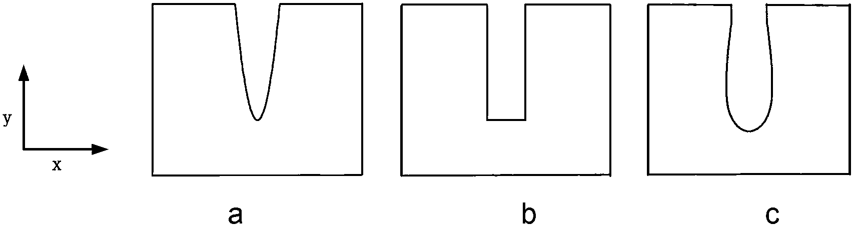
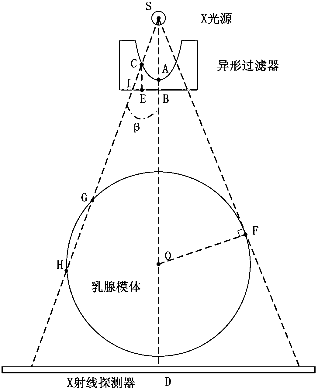
[0024] In the process of using X-ray machines and X-ray detectors for mammography, since the cross-section of the mammary phantom is close to an elliptical shape, the attenuation path lengths of X-rays in the mammary phantom are different. The X-ray attenuation is larger at the position, and the X-ray attenuation is smaller at the position away from the center of the mammary phantom, thus resulting in a larger range of the number of photons received...


PUM


Abstract

The invention discloses an imaging method used for X-radiography. The imaging method comprises the following steps of: setting a special shape aluminum filter at the central position of a light source of an X-ray machine when an X-ray machine and an X-ray detector are utilized to carry out mammography, absorbing low-energy photons sent by the X-ray machine by the special shape aluminum filter, reducing the quantity scope of received photons on the X-ray detector, and adjusting the X-ray intensity and the regional quantity of different positions of a breast model region, wherein the thickness of the special shape aluminum filter is gradually increased from the center to the external edge. The imaging method can reduce the quantity scope of the received photons on the X-ray detector, adjust the X-ray intensity and the regional quantity, and enhance the imaging quality.

Description

technical field [0001] The invention relates to the technical field of X-ray imaging, in particular to an imaging method for X-ray photography. Background technique [0002] At present, in mammary X-ray imaging techniques, such as mammography or breast-specific CT imaging, X-ray machines and X-ray detectors are used to perform two-dimensional or three-dimensional imaging of breast structures. In this configuration, the patient's breast is placed in the cone beam emitted by the X-ray machine, and the X-ray detector is used to capture the X-ray beam passing through the patient's breast. [0003] Breast X-ray imaging technology only uses mammary gland receptors as the detection object. According to the regular structure of mammary gland receptors, the X-ray intensity after passing through mammary gland receptors and reaching the detector has certain characteristics. The cross-section of mammary gland receptors is close to an elliptical shape. , and the attenuation path lengths...

Claims


Application Information

Patent Timeline
IPC IPC(8): A61B6/00
Inventor 王哲赵维魏存峰王燕芳舒航魏龙
Owner INST OF HIGH ENERGY PHYSICS CHINESE ACADEMY OF SCI
Why Eureka
Try Eureka