Adaptive hierarchical chest large data display implementation method

An implementation method and layered technology, applied in the field of medical image processing, can solve problems such as inability to satisfy clear rendering, inability to obtain three-dimensional rendering results, and ordinary computers unable to load large amounts of data calculations, etc.

Inactive Publication Date: 2013-05-01
HEBEI UNIVERSITY
View PDF2 Cites 11 Cited by
  • Summary
  • Abstract
  • Description
  • Claims
  • Application Information

AI Technical Summary

Problems solved by technology

First of all, the increase in the amount of data has caused ordinary computers to be unable to load calculations with large amounts of data, and cannot o...

Method used


Image

  • Adaptive hierarchical chest large data display implementation method
  • Adaptive hierarchical chest large data display implementation method
  • Adaptive hierarchical chest large data display implementation method

Examples


Embodiment

[0055] This embodiment is realized in a computer with Pentium® Dual-Core CPU E5800 3.20GHz, graphics card is NVIDIA GeForce GT 430, internal memory is 2.0GB, operating system is Window XP, and the whole hybrid rendering method is written in c++ language.

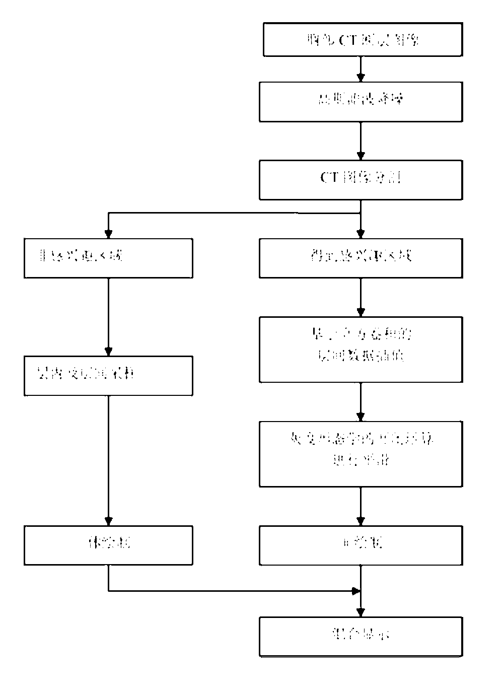
[0056] The implementation process of this embodiment is as follows figure 1 shown.

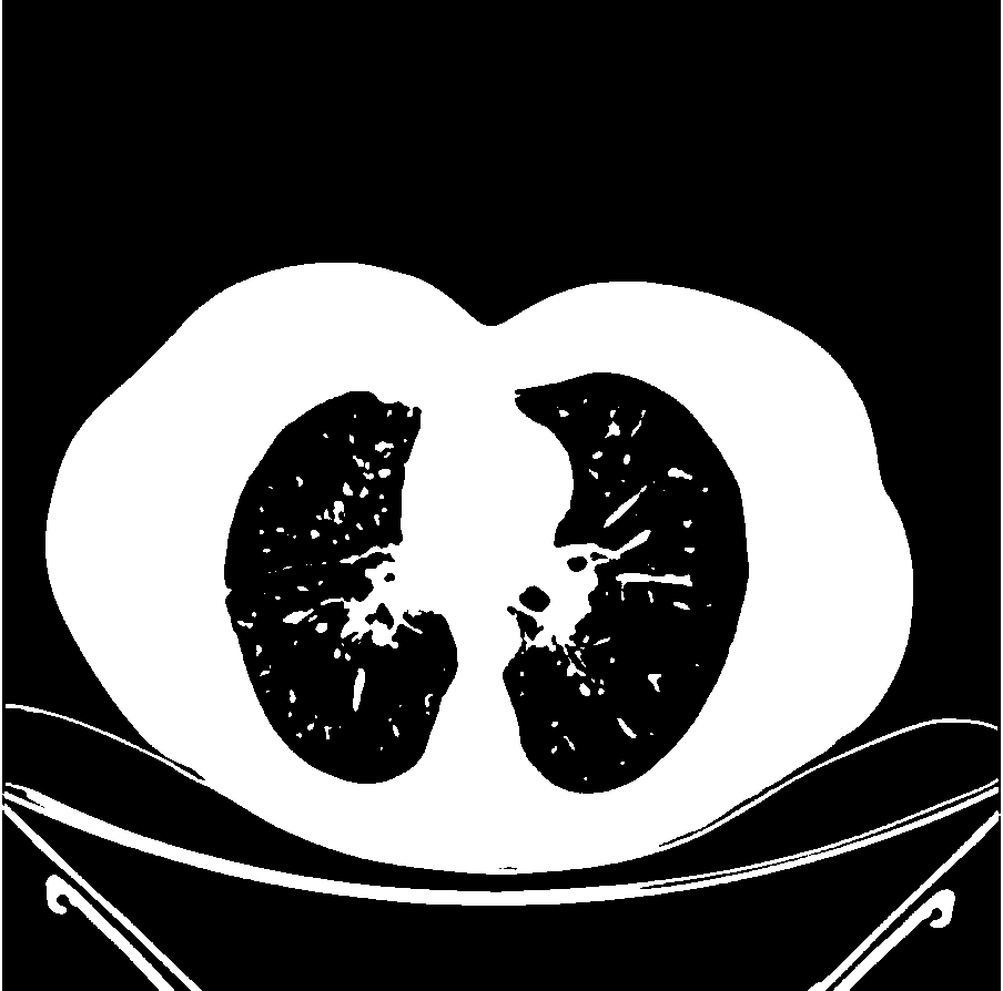
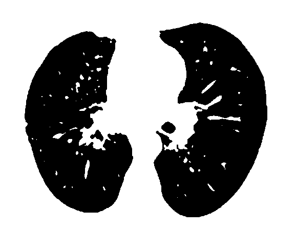
[0057] In the first step, the original CT image sequence of the chest ( figure 2 Shown is one of the sequence images) Gaussian filter noise reduction method is used for noise reduction processing, and the region growth algorithm is used for region growth in the denoised chest CT image, and different tag values ​​are assigned to each part, and the original sequence is saved to form Original image sequence; select the marker value corresponding to the region of interest (here, the lungs), extract the corresponding CT values ​​(gray information) of all pixels corresponding to the marker value in the original image, press The original image s...


PUM


Abstract

The invention discloses an adaptive hierarchical chest large data display implementation method, which comprises the following steps: dividing a CT (computed tomography) sectional image into a target layer image sequence and a background layer image sequence; performing interlayer cubic convolution interpolation on the divided target layer image sequence to form volume data of which each axial resolution is appropriate; making a clear surface drawing by a marching cubes algorithm; performing intraformational and interlayer sampling on the data of the background layer sequence to obtain uninteresting region data of which the resolution is reduced; making a rough body drawing for the data by a ray casting algorithm; displaying the drawing results of the clear surface drawing and the rough body drawing at the same time, and performing hybrid three-dimensional visualization. By the method, the local resolution in the interesting region is increased, and the anisotropic resolution of the uninteresting region is reduced, so that a user is facilitated to know the structure information of a local interesting region, the data volume is decreased and drawing can be implemented on an ordinary computer.

Description

technical field [0001] The present invention relates to a realization method of self-adaptive layered chest big data display, especially to the image segmentation of the chest cavity and lungs, the realization method of interlayer interpolation and intralayer sampling in the designated area and the realization method of chest partition visualization, which belongs to medical image processing technology field. Background technique [0002] The three-dimensional reconstruction and visualization of medical images is to use the characteristics of the human visual system to interpolate the two-dimensional discrete data generated by medical imaging equipment on the computer, and convert them into intuitive, three-dimensional images for displaying the three-dimensional shape of tissues and organs. Structural information that cannot be obtained by traditional means. 3D reconstruction and visualization have changed the traditional way of reading films, can provide doctors with anato...

Claims


Application Information

Patent Timeline
IPC IPC(8): G06T17/00G06T7/00
Inventor 顾力栩王兵樊星董斌
Owner HEBEI UNIVERSITY
Why Eureka
Try Eureka