Method for segmenting aortic-valve ultrasound image sequence based on interframe-shape-constraint GCV model

An aortic valve and ultrasound image technology, which is applied in the field of medical ultrasound image segmentation, and can solve the problems of severe overflow and incomplete segmentation of aortic valve ultrasound images.

Active Publication Date: 2014-02-26
HEBEI UNIVERSITY
View PDF8 Cites 5 Cited by
  • Summary
  • Abstract
  • Description
  • Claims
  • Application Information

AI Technical Summary

Problems solved by technology

[0004] The purpose of the present invention is to provide a method for segmenting aortic valve ultrasonic image sequences based on inter-frame shape constraints of th...

Method used


Image

  • Method for segmenting aortic-valve ultrasound image sequence based on interframe-shape-constraint GCV model
  • Method for segmenting aortic-valve ultrasound image sequence based on interframe-shape-constraint GCV model
  • Method for segmenting aortic-valve ultrasound image sequence based on interframe-shape-constraint GCV model

Examples


Embodiment Construction

[0035] The present invention will be further described below in conjunction with the accompanying drawings and a specific example.

[0036] This example is in Dual-Core CPU E5800 3.20GHz, graphics card is NVIDIA GeForce GT430NVIDIA GeForce GT430, memory is 2.00GB, the operating system is Window XP, and the whole segmentation method is written in C++ and Matlab language.

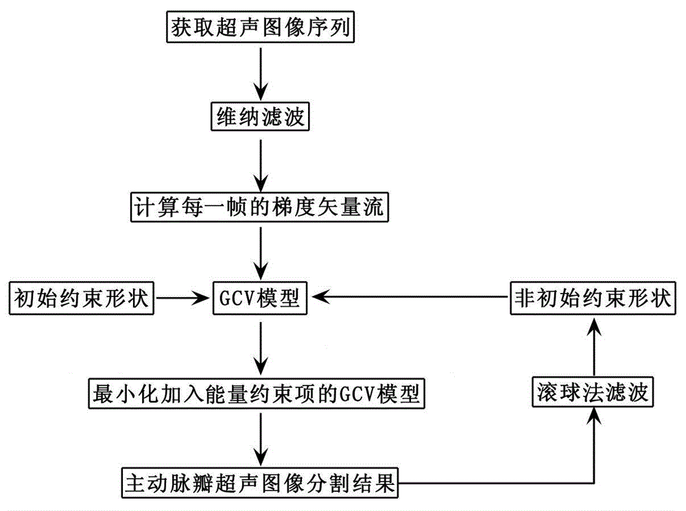
[0037] The procedure of this method is as follows figure 1 The steps shown are performed:

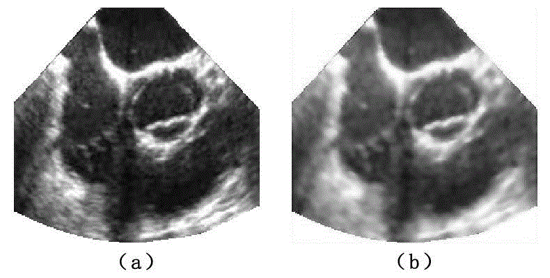
[0038] (1) Acquire a set of continuous ultrasound image sequences of the aortic valve, and extract the fan-shaped area of ​​each frame, such as figure 2 (a), the threshold of the non-sector area is 255; then Wiener filtering is performed on the image acquired in each frame, and the filtered result is as follows figure 2 (b).

[0039] After the Wiener filtering process, the speckle noise in the ultrasound image can be removed, and the edge information can be well preserved.

[0040] (2) Build the GCV model:

[...


PUM


Abstract

The invention discloses a method for segmenting an aortic-valve ultrasound image sequence based on an interframe-shape-constraint GCV model. The method includes the following steps: acquiring a group of ultrasound images and carrying out Wiener filtering; calculating a gradient vector flow field of each image frame by frame and adding the gradient vector flow fields to a CV model as energy constraints and obtaining the GCV model; through defining an initial constraint shape and adding the initial constraint shape into the GCV model as an energy constraint item and then minimizing an energy functional so that a segmentation result of a first frame of image is obtained; and carrying out filtering of a rolling sphere method on the aortic-valve segmentation result of an adjacent previous-frame image and adding the obtained result into the GCV model as an energy constraint item so that the segmentation result of a current frame is obtained through calculation. The method for segmenting the aortic-valve ultrasound image sequence based on the interframe-shape-constraint GCV model is operated on short-axis images of ultrasonic cardiograms so that not only is work load of doctors reduced, a problem that serious overflow in segmentation of the aortic-valve ultrasound images in the prior art is also solved. The segmentation result is extremely close to the result of manual segmentation. The method enables the aortic valve to be segmented in a simple and highly-efficient manner.

Description

technical field [0001] The invention relates to a medical ultrasonic image segmentation method, in particular to a method for segmenting an aortic valve ultrasonic image sequence based on a GCV model constrained by inter-frame shapes. Background technique [0002] In my country, aortic valve disease is the most common, complex and dangerous cardiovascular disease, which seriously endangers human health. The aortic valve is located at the junction of the end of the left ventricular outflow tract and the aorta. It acts like a "one-way valve" to prevent blood backflow and ensure the normal and good pumping function of the heart. Due to the safe, non-invasive, simple, cheap, and repeatable characteristics of B-ultrasound, echocardiography has been widely used in clinical diagnosis to observe the shape and position of the aortic valve. The accurate segmentation of the aortic valve in the ultrasound image can not only assist doctors in clinical diagnosis, but also the basis of im...

Claims


Application Information

Patent Timeline
IPC IPC(8): G06T7/00G06T5/00A61B8/00
Inventor 顾力栩董斌郭怡婷王兵
Owner HEBEI UNIVERSITY
Why Eureka
Try Eureka