Medical image processing method

A processing method and medical image technology, applied in image data processing, image analysis, application, etc., can solve the problems that are not conducive to the popularization and promotion of neurointerventional surgery, reduce blood flow velocity, and cannot simultaneously develop the proximal and distal ends of the occluded segment, etc. question

Inactive Publication Date: 2014-05-21
姜卫剑
View PDF0 Cites 11 Cited by
  • Summary
  • Abstract
  • Description
  • Claims
  • Application Information

AI Technical Summary

Benefits of technology

This new technology uses different techniques such as combining magnetic resonance imaging (MRI), DSA or CT scans together into one composite picture called an enhanced view showing both arteries and veins near their blockage location. These pictures help healthcare professionals see how they have been blocked by plaque buildup over time better understanding what caused damage during treatment procedures.

Problems solved by technology

Technicians have been unable to accurately detect if there were any abrupt changes in the heart caused by strokes due to blockages like clots within blood vessels. Current medical procedures involve invasiveness, making them difficult to apply widely across all types of stroke cases without causing complications. There currently exist noninvasive therapy options including ultrasound waves, X ray fluoroscopy, laser Doppler angioangraphy, opthalmology, etc., while current technologies require multiple sessions before accurate results may be obtained.

Method used


Image

  • Medical image processing method
  • Medical image processing method
  • Medical image processing method

Examples


Embodiment Construction

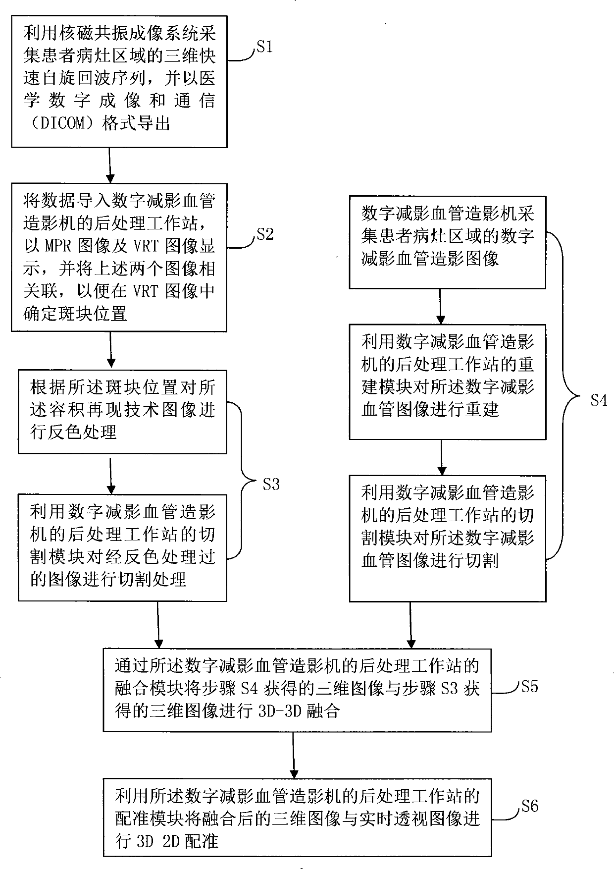
[0027] Referring to the accompanying drawings, the specific implementation of the medical image processing method of the present invention will be described in detail.

[0028] The nuclear magnetic resonance system described in the present invention is a MAGNETOM Skyra3.0T intelligent magnetic resonance imager produced by German Siemens.

[0029] The digital subtraction angiography machine (3D-DSA) described in the present invention is the large plate blood vessel machine that the model of German Siemens Company is Artis Zeego.

[0030] The post-processing workstation of the digital subtraction angiography machine described in the present invention is a CT medical imaging workstation, which is an imaging diagnostic workstation for DICOM and non-DICOM CT equipment, integrating image transmission or collection, image reading, processing, and reporting , and provide professional work modules such as image reconstruction, image cutting, image fusion, and image registration. For e...


PUM


Abstract

The invention relates to a medical image processing method which is characterized in that images formed by three-dimensional fast spin echo imaging collected by a magnetic resonance imaging system in the focus area of a patient and blood vessel images collected by a digital subtraction angiography machine are subjected to reconstruction, color inverting, cutting, 3D-3D fusion and 3D-2D registering through a post-processing work station of the digital subtraction angiography machine, so that three-dimensional blood vessel graphs can be overlapped on a real-time radio-fluoroscopic image. Therefore, by adopting the medical image processing method, real-time imaging guide can be performed during nerve involvement surgeries for blocked blood vessels, the near end and far end of the closed section of the blood vessel can be shown simultaneously through imaging, and physicians can clearly get known of conditions in the focus areas and further can perform operating better.

Description


Claims


Application Information

Patent Timeline
Owner 姜卫剑
Why Eureka
Try Eureka