Automatic breast ultrasound scanning method

A breast and ultrasound technology, applied in ultrasound/sonic/infrasonic Permian technology, ultrasound/sonic/infrasound image/data processing, organ motion/change detection, etc. Check for accuracy, etc.

Active Publication Date: 2014-10-15
SHANTOU INST OF UITRASONIC INSTR CO LTD
View PDF6 Cites 19 Cited by
  • Summary
  • Abstract
  • Description
  • Claims
  • Application Information

AI Technical Summary

Problems solved by technology

Although this method can realize the automatic scanning of the mammary gland, it is beneficial to improve the inspection efficiency, but the person under inspection must adopt a prone position during the inspection process, which feels uncomfortable, and the probe moving device and the probe are immersed in water, and the probe moves The de

Method used


Image

  • Automatic breast ultrasound scanning method
  • Automatic breast ultrasound scanning method
  • Automatic breast ultrasound scanning method

Examples


Embodiment 1

[0039] The breast ultrasound automatic scanning method of this embodiment uses a breast ultrasound automatic scanning device to scan the examinee's breasts.

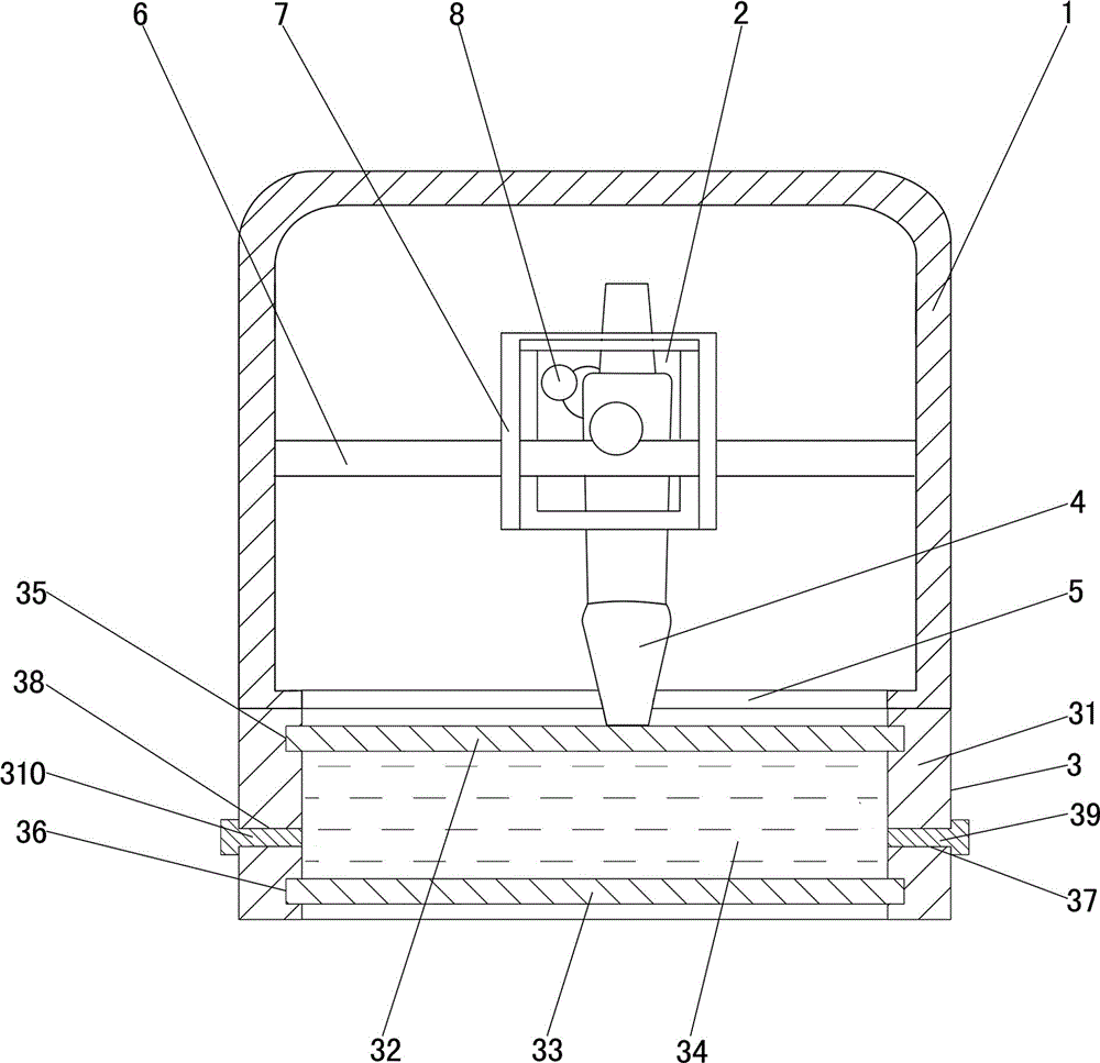
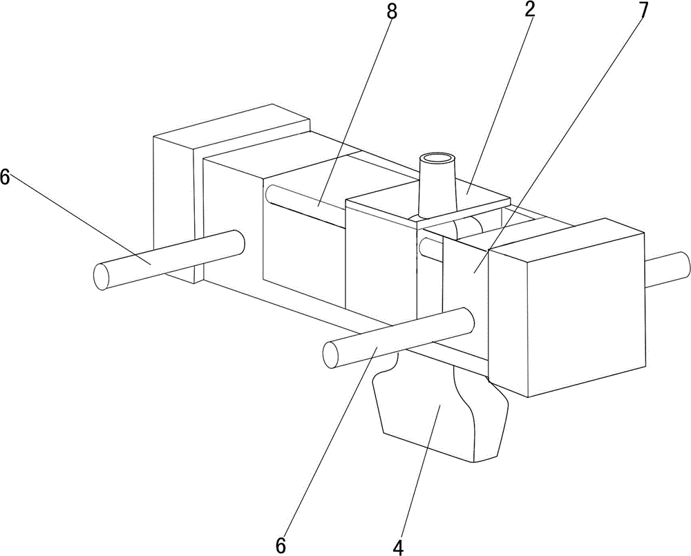
[0040] Such as figure 1 As shown, the breast ultrasound automatic scanning device used includes a housing 1, an ultrasonic coupling device 3, a probe base 2, an ultrasonic probe 4 installed on the probe base 2, and a probe that can drive the probe base 2 and the ultrasonic probe 4 to move together. device. The probe seat 2, the ultrasonic probe 4 and the probe moving device are arranged in the casing 1, and the bottom of the casing 1 is provided with an opening 5, and the ultrasonic coupling device 3 is installed on the opening 5 at the bottom of the casing 1; the ultrasonic coupling device 3 includes a support frame 31, an upper The elastic membrane 32 and the lower elastic membrane 33, the supporting frame body 31 is connected with the edge of the opening 5 at the bottom of the casing, the edges of the upper elastic m...

Embodiment 2

[0059] The breast ultrasound automatic scanning method of this embodiment uses a breast ultrasound automatic scanning device to scan the examinee's breasts.

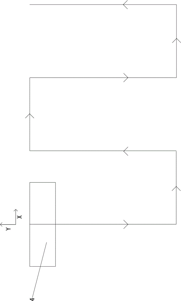
[0060] Such as Figure 4 As shown, in the breast ultrasound automatic scanning device used in the present embodiment, a couplant tank 9 is also provided in the housing 1, an opening 10 is provided at the upper end of the couplant tank 9, and an opening 10 that can drive the ultrasonic probe 4 is provided on the probe base 2. Ultrasonic probe lifting mechanism. The coupling agent groove 9 is arranged at a position corresponding to the starting end of the moving line of the ultrasonic probe. A couplant is contained in the couplant tank 9 .

[0061] In this embodiment, the ultrasonic probe lifting mechanism includes a lifting motor and a transmission mechanism. The lifting motor drives the ultrasonic probe to lift through the transmission mechanism; the transmission mechanism includes a lifting screw 11 and a lifting nut ...


PUM


Abstract

Disclosed is an automatic breast ultrasound scanning method. The method is characterized in that a used automatic breast ultrasound scanning device comprises an ultrasonic coupling device, a probe seat, an ultrasonic probe and a probe moving device; the ultrasonic coupling device comprises a supporting frame, an upper elastic membrane and a lower elastic membrane, the edges of the upper elastic membrane and the lower elastic membrane are both connected with the supporting frame, and an airtight cavity is formed between the upper elastic membrane and the lower elastic membrane and is filled with coupling liquid; when scanning is performed, an examined person lies on an examination couch on the back, then the automatic breast ultrasound scanning device is put on the breast of the examined person, and the lower elastic membrane of the ultrasonic coupling device is tightly pressed on the breast; then the probe moving device drives the probe seat and the ultrasonic probe to move together according to a predetermined route, and the ultrasonic probe clings to the upper surface of the upper elastic membrane and scans the breast. The breast can be scanned automatically; the examined person adopts a supine posture in the process of examination, thereby feeling comfortable; operation is stable, scanning speed is high, and examination accuracy is high.

Description

technical field [0001] The invention relates to a method for detecting human tissue by ultrasonic waves, in particular to an automatic ultrasonic scanning method for mammary glands. Background technique [0002] Existing breast ultrasound examinations are generally manual scans, which have high requirements on the doctor's scanning techniques during the breast examination process, and the scanning speed is slow, which affects the examination efficiency. [0003] The emergence of breast ultrasound automatic scanning device helps to improve the scanning speed and inspection efficiency. An existing breast ultrasound automatic scanning device includes a mammary gland examination bed, a probe and a probe moving device for the subject to be examined in a prone position; two inspection holes are arranged in the chest area of ​​the breast examination bed; the probe is fixedly installed The probe moving device is connected with the ultrasonic diagnostic instrument; a water tank is a...

Claims


Application Information

Patent Timeline
IPC IPC(8): A61B8/08
Inventor 李德来蔡泽杭郭境峰
Owner SHANTOU INST OF UITRASONIC INSTR CO LTD
Why Eureka
Try Eureka