Primary skin color keeping vein development method

A vein imaging and primary color technology, applied in the field of medical image processing, can solve the problems of inconspicuous detail features and color distortion.

Active Publication Date: 2014-10-29
GUANGZHOU INST OF ADVANCED TECH CHINESE ACAD OF SCI
View PDF3 Cites 14 Cited by
  • Summary
  • Abstract
  • Description
  • Claims
  • Application Information

AI Technical Summary

Problems solved by technology

However, when traditional image fusion algorithms are applied to the display of v

Method used


Image

  • Primary skin color keeping vein development method
  • Primary skin color keeping vein development method
  • Primary skin color keeping vein development method

Examples


no. 1 example

[0104]The vein imaging device of the present invention is used to display vein images on a display that can clearly and accurately display vein distribution and maintain the original color of non-vein areas, and includes a display 1 and an image processing module 2 .

[0105] And image processing module 2 includes:

[0106] The digital image processing chip is used to complete the specific implementation process of a vein imaging method that maintains the original color of the epidermis according to the present invention. The input end is connected to the visible light image acquisition module and the near-infrared image acquisition module, and the output end is used to display the processed results. The connected display 1;

[0107] The matching identifier 5 placed on the upper right corner of the target object 7 is used for feature identification during image registration.

[0108] The target object 7 is placed in the middle of the substrate 6 and within the imaging range o...

Embodiment 2

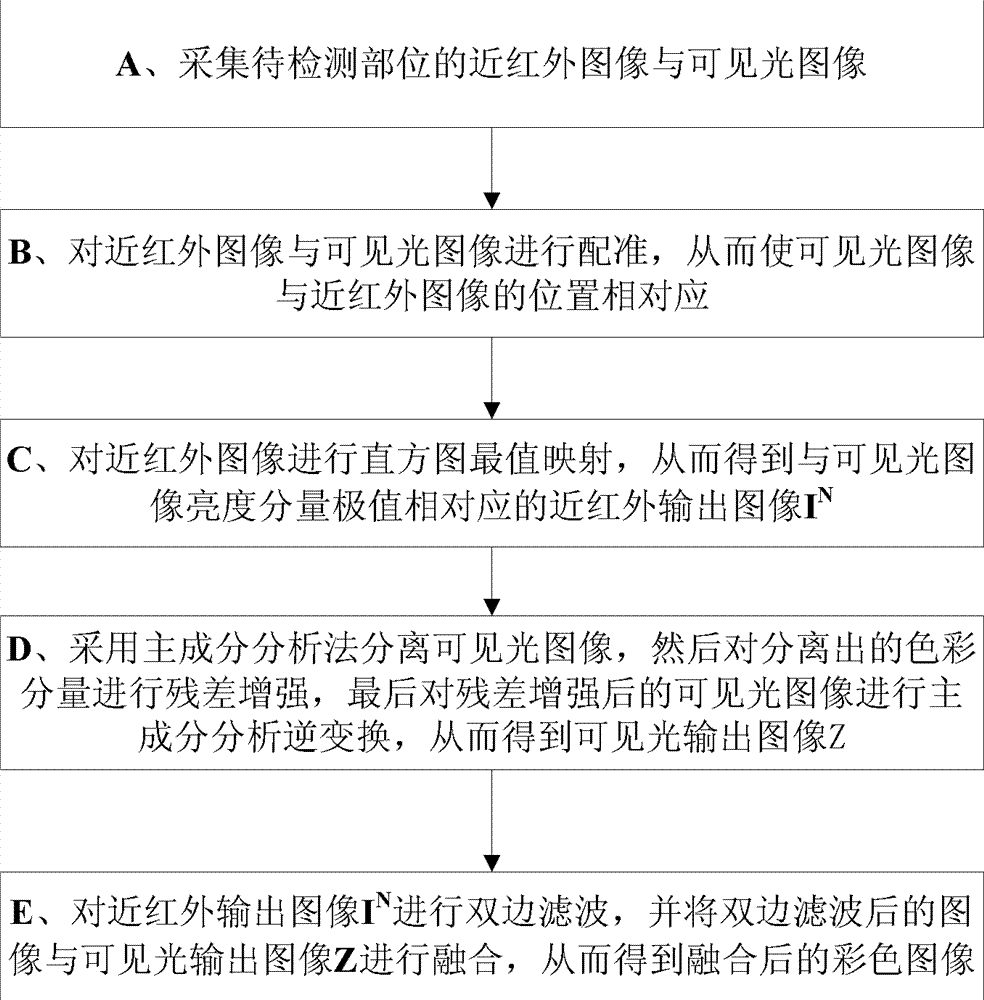
[0119] The present invention proposes a real-time image fusion method based on histogram maximum value mapping, principal component analysis and bilateral filtering, which is used for fusion of near-infrared and color images in vivo or in vitro. The method has a fast processing speed, so that an image including the distribution of subepidermal veins and maintaining the original color of the epidermis can be obtained in real time, quickly and efficiently.

[0120] The concrete realization process of a kind of vein imaging method that keeps epidermis original color of the present invention is as follows:

[0121] Step S1: collecting near-infrared and visible light images of the part to be detected;

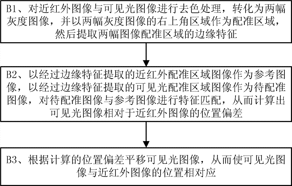
[0122] Step S2: Perform image registration according to the near-infrared image and the color image acquired in the same scene;

[0123] The image registration process can be further divided into:

[0124] S21. Mark a special symbol (such as a cross) in the upper right corner of t...


PUM


Abstract

The invention discloses a primary skin color keeping vein development method. The primary skin color keeping vein development method comprises the steps of acquiring a near infrared image and a visible light image at a position to be detected; matching the near infrared image with the visible light image; performing histogram most value mapping on the near infrared image and obtaining a near infrared output image corresponding to a visible light image luminance component extreme value; adopting a principal component analysis method to separate the visible light image, then performing residual error strengthening on a separated color component and finally performing principal component analysis inverse transformation on the visible light image subjected to the residual error strengthening; performing bilateral filtering on the near infrared output image and merging the image obtained after bilateral filtering with the near infrared output image Z to obtain a merged color imaged. By means of the primary skin color keeping vein development method, the problem that a traditional vein display image is unreal is solved, the merged color image can be ensured to be accurate in vein distribution and obvious in details, and the primary skin color in a non-vein region can be kept. The primary skin color keeping vein development method can widely applied to the field of medical image processing.

Description

technical field [0001] The invention relates to the field of medical image processing, in particular to a vein imaging method that maintains the original color of the epidermis. Background technique [0002] The acquisition and processing of vein distribution images has important application value in the fields of individual recognition and medical diagnosis and treatment, and has become one of the important research topics in the field of biomedical imaging. Since it is often difficult to obtain complete vein distribution information in the visible light band, people usually use the strong absorption characteristics of hemoglobin for near-infrared light to obtain clear vein distribution maps through infrared imaging. The use of vein display equipment for children's venipuncture can significantly improve the success rate of puncture. [0003] At present, there are mainly two ways to obtain subcutaneous vein distribution maps: vein display and vein projection: vein display u...

Claims


Application Information

Patent Timeline
IPC IPC(8): G06T5/00G06T5/50
Inventor 邸思刘鹏金建陈贤帅杜如虚
Owner GUANGZHOU INST OF ADVANCED TECH CHINESE ACAD OF SCI
Why Eureka
Try Eureka