Method and system for rapid and automatic acquisition of vessel edge morphology in dynamic ultrasound images

An ultrasonic image and automatic acquisition technology, applied in the field of medical electronic information, can solve the problems of difficult processing algorithm and low system efficiency.

Active Publication Date: 2017-02-01
YUNNAN UNIV +1
View PDF8 Cites 0 Cited by
  • Summary
  • Abstract
  • Description
  • Claims
  • Application Information

AI Technical Summary

Problems solved by technology

The problem encountered in the continuous testing and application in the later stage is that the efficiency of the system is not high, and it can only process 3 to 6 frames of images per second
In the case of real-time processing with high quality requirements and acquisition of 20-30 frames per second, complex processing algorithms have encountered difficulties

Method used


Image

  • Method and system for rapid and automatic acquisition of vessel edge morphology in dynamic ultrasound images
  • Method and system for rapid and automatic acquisition of vessel edge morphology in dynamic ultrasound images
  • Method and system for rapid and automatic acquisition of vessel edge morphology in dynamic ultrasound images

Examples


Embodiment Construction

[0078] The implementation technical scheme of the method and system for fast and automatic acquisition of blood vessel edge morphology in dynamic ultrasonic images of the present invention will be described in detail and completely below in conjunction with the accompanying drawings.

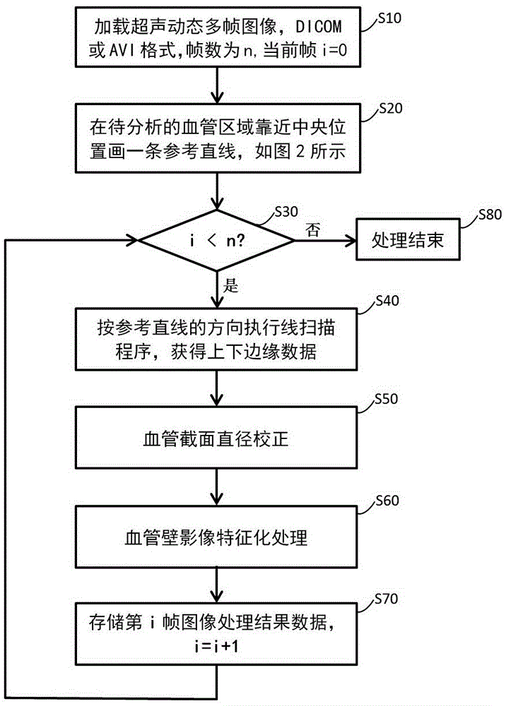
[0079] Such as figure 1 As shown, the method for rapid and automatic acquisition of vessel edge morphology in dynamic ultrasound images includes the following steps:

[0080] Step S10 , loading multiple frames of vascular ultrasonic dynamic images, obtaining the total image frame number ImageFrame_n from a file in DICOM or AVI format, and reading each frame of image. The image processed by the method of the invention only uses grayscale data, so the image can be decolorized and the grayscale can be retained. If Bits Allocated>8 and Bit Stored>8 for each pixel of the DICOM image, convert the grayscale value of each image pixel to 8 bits, 256 grayscale levels. In terms of processing effect, the ...


PUM


Abstract

The invention discloses a method and system for rapidly and automatically collecting blood vessel edge forms in a dynamic ultrasonic image and belongs to the field of medical electronic information. The method mainly includes the steps of extracting blood vessel edge vector data in the ultrasonic image of a line scanning mode and calculating and correcting blood vessel inner diameters. According to the processing process of the method, complex arithmetical operations such as image preprocessing, edge detecting and vectorizing in traditional edge detecting are converted into a simple detecting method operated based on numerical value addition and subtraction methods. Compared with a traditional method, the processing speed of the method and system for rapidly and automatically collecting the blood vessel edge forms in the dynamic ultrasonic image can be improved by multiple times even ten or more times, and the real-time detecting and processing requirement can be met in dynamic blood vessel ultrasonic detecting of 20 or more frames per second. The data quality of processing results meets the requirement for later-stage analyzing and diagnosis references through finite experiments.

Description

technical field [0001] The invention belongs to the field of medical electronic information, and relates to image edge extraction technology in digital image processing, in particular to a method and system for quickly and automatically collecting blood vessel edge shapes in dynamic ultrasonic images. Background technique [0002] Cardiovascular disease is one of the biggest health killers of human beings in this century, and ultrasonic non-destructive detection is the most commonly used and reliable method at present. With the improvement of people's quality of life and attention to their own health, the number of patients who require cardiovascular status checks has increased significantly, making manual measurement of some key indicators of vascular imaging more and more burdensome. [0003] References "Development and Application of Auto-Tracing Vessel Image Analysis System (Development and Application of Auto-Tracing Vessel Image Analysis System), Journal of Kunming Med...

Claims


Application Information

Patent Timeline
Patent Type & Authority Patents(China)
IPC IPC(8): G06T7/00G06T5/00
Inventor 郑家亮丁云川雷晓凌
Owner YUNNAN UNIV
Why Eureka
Try Eureka